. 2010 Dec 28;107(52):22629-34.
doi: 10.1073/pnas.1012141108. Epub 2010 Dec 13.

Follicle-stimulating hormone increases bone mass in female mice

Affiliations

Follicle-stimulating hormone increases bone mass in female mice

Charles M Allan et al. Proc Natl Acad Sci U S A. .

Abstract

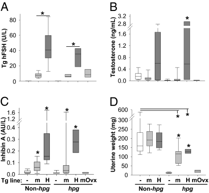
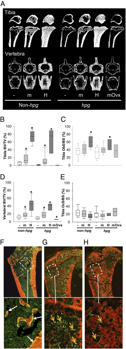
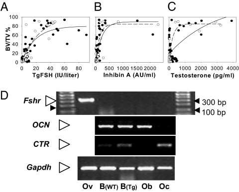
Elevated follicle-stimulating hormone (FSH) activity is proposed to directly cause bone loss independent of estradiol deficiency in aging women. Using transgenic female mice expressing human FSH (TgFSH), we now reveal that TgFSH dose-dependently increased bone mass, markedly elevating tibial and vertebral trabecular bone volume. Furthermore, TgFSH stimulated a striking accrual of bone mass in hypogonadal mice lacking endogenous FSH and luteinizing hormone (LH) function, showing that FSH-induced bone mass occurred independently of background LH or estradiol levels. Higher TgFSH levels increased osteoblast surfaces in trabecular bone and stimulated de novo bone formation, filling marrow spaces with woven rather than lamellar bone, reflective of a strong anabolic stimulus. Trabecular bone volume correlated positively with ovarian-derived serum inhibin A or testosterone levels in TgFSH mice, and ovariectomy abolished TgFSH-induced bone formation, proving that FSH effects on bone require an ovary-dependent pathway. No detectable FSH receptor mRNA in mouse bone or cultured osteoblasts or osteoclasts indicated that FSH did not directly stimulate bone. Therefore, contrary to proposed FSH-induced bone loss, our findings demonstrate that FSH has dose-dependent anabolic effects on bone via an ovary-dependent mechanism, which is independent of LH activity, and does not involve direct FSH actions on bone cells.


Conflict of interest statement

The authors declare no conflict of interest.

Figures

Fig. 1.
Fig. 1.
TgFSH-induced changes to serum hormones and uterine weights. Groups comprised adult females from TgFSHm (m) or TgFSHH (H) lines and non-Tg (−) controls with non-hpg or hpg backgrounds (n = 7–26 mice per group). TgFSHm hpg females were ovariectomized (Ovx) at 3 mo old (n = 7). Box plots show quartile values as box boundaries with median indicated by enclosed horizontal line and whiskers indicating 5th and 95th percentiles when definable (≥9 per group). As expected, serum TgFSH levels were up to 5-fold higher (P < 0.01) in TgFSHH versus TgFSHm females (A). TgFSH elevated (P < 0.001) serum testosterone (B) and inhibin A (C) in TgFSHH hpg females compared with non-Tg controls. Compared with WT values, uterine weights from TgFSHm or TgFSHH non-hpg females were equivalent (D), whereas those from TgFSHm hpg or TgFSHH hpg females were lower (P < 0.01). Asterisk indicates significant differences (P < 0.05) between TgFSH and respective control groups or comparisons indicated by lines above.
Fig. 2.
Fig. 2.
TgFSH increased bone mass in adult female mice. (A) Representative longitudinal and cross-sections of proximal tibia taken 0.5 mm below growth plate and middle of L3 vertebra (μCT) are shown. Histomorphometry used bones from all collected TgFSHm (n = 26 non-hpg or 16 hpg), TgFSHH (n = 15 non-hpg or 8 hpg), TgFSHm hpg-Ovx (n = 7), and non-Tg (n = 19 non-hpg or 18 hpg) females. (B–E) Box plots show quartile values as box boundaries with median indicated by enclosed horizontal line and whiskers indicating 5th and 95th percentiles when definable (≥9 per group). TgFSH increased trabecular BV/TV (μCT) in tibia (P < 0.01) of TgFSH females (B) and vertebra (P < 0.001) of TgFSHH females (D, non-hpg or hpg) versus respective non-Tg controls. Asterisk indicates significant differences (P < 0.05) between TgFSH and respective control groups. Tibial trabecular Ob.S/BS or Oc.S/BS (static histomorphometry) show a trend for increased Ob.S/BS and normal Oc.S/BS in TgFSH versus non-Tg females (C and E). (F–H) Calcein (green) and xylenol orange staining show normal bone in WT with expected calcein double labels (F, white arrows) compared with bone in TgFSHH females exhibiting marrow spaces actively being filled with woven bone as indicated by extensive diffuse calcein uptake (G, orange arrows) or almost completely filled by woven bone with little ongoing formation as indicated by reduced calcein uptake (H, orange arrows). (Magnification: Upper, 40×; Lower, 200×.)
Fig. 3.
Fig. 3.
Increased bone mass correlated with serum hormone levels in TgFSH females. (A–C) Scatter plots and correlation lines (Sigmaplot version 10 Wizard Correlation) for tibial trabecular BV/TV (%) versus serum TgFSH (A) or ovarian-derived serum inhibin A (B) or testosterone levels (C) shown as solid lines and filled circles for non-hpg data or dashed lines and open circles for hpg data. Data were combined for TgFSHm and TgFSHH lines on either the non-hpg (FSH, n = 54; inhibin A, n = 42; testosterone, n = 57) or hpg (FSH, n = 32; inhibin A, n = 34; testosterone, n = 41) background. Correlations indicated an exponential rise to maximal BV/TV associated with increasing levels of all serum hormones measured. (D) Mouse Fshr mRNA expression was detected (RT-PCR) in adult ovary (Ov) as expected but not in long bone from either TgFSHH (Tg) or non-Tg (WT) adult females or in isolated cultured mouse osteoblast (Ob) or osteoclast (Oc) preparations. CTR or OCN mRNA expression confirmed differentiated Oc or Ob cells, respectively. Gapdh mRNA was used as internal cDNA control, and Fshr findings repeated in triplicate.

References

    1. Simoni M, Gromoll J, Nieschlag E. The follicle-stimulating hormone receptor: Biochemistry, molecular biology, physiology, and pathophysiology. Endocr Rev. 1997;18:739–773. - PubMed
    1. Lenton EA, Sexton L, Lee S, Cooke ID. Progressive changes in LH and FSH and LH: FSH ratio in women throughout reproductive life. Maturitas. 1988;10:35–43. - PubMed
    1. Robertson DM, Hale GE, Fraser IS, Hughes CL, Burger HG. A proposed classification system for menstrual cycles in the menopause transition based on changes in serum hormone profiles. Menopause. 2008;15:1139–1144. - PubMed
    1. Sherman BM, Korenman SG. Hormonal characteristics of the human menstrual cycle throughout reproductive life. J Clin Invest. 1975;55:699–706. - PMC - PubMed
    1. Klein NA, et al. Decreased inhibin B secretion is associated with the monotropic FSH rise in older, ovulatory women: A study of serum and follicular fluid levels of dimeric inhibin A and B in spontaneous menstrual cycles. J Clin Endocrinol Metab. 1996;81:2742–2745. - PubMed

Publication types

MeSH terms