. 2010 Dec 28;107(52):22593-8.
doi: 10.1073/pnas.1011624108. Epub 2010 Dec 13.

T-cell cytokines differentially control human monocyte antimicrobial responses by regulating vitamin D metabolism

Affiliations

T-cell cytokines differentially control human monocyte antimicrobial responses by regulating vitamin D metabolism

Kristina Edfeldt et al. Proc Natl Acad Sci U S A. .

Abstract

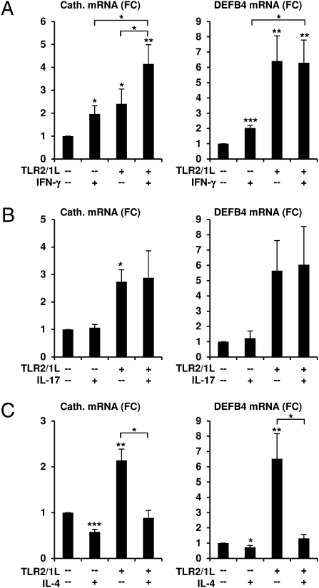
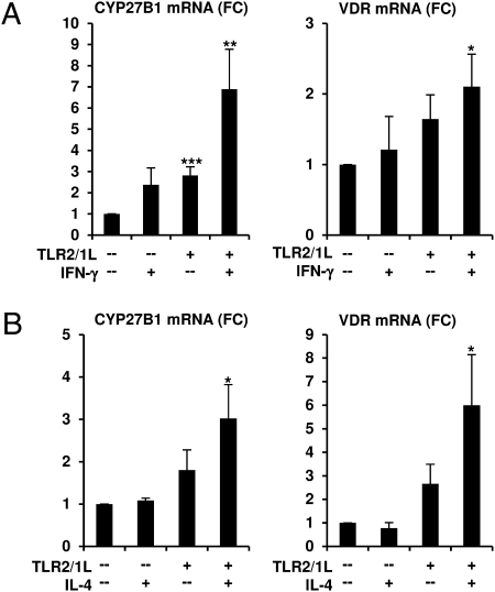
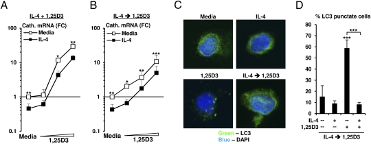
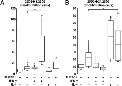
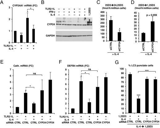
We investigated the mechanisms by which T-cell cytokines are able to influence the Toll-like receptor (TLR)-induced, vitamin D-dependent antimicrobial pathway in human monocytes. T-cell cytokines differentially influenced TLR2/1-induced expression of the antimicrobial peptides cathelicidin and DEFB4, being up-regulated by IFN-γ, down-regulated by IL-4, and unaffected by IL-17. The Th1 cytokine IFN-γ up-regulated TLR2/1 induction of 25-hydroxyvitamin D-1α-hydroxylase (i.e., CYP27B1), leading to enhanced bioconversion of 25-hydroxyvitamin D(3) (25D(3)) to its active metabolite 1,25D(3). In contrast, the Th2 cytokine IL-4, by itself and in combination with the TLR2/1 ligand, induced catabolism of 25D(3) to the inactive metabolite 24,25D(3), and was dependent on expression of vitamin D-24-hydroxylase (i.e., CYP24A1). Therefore, the ability of T-cell cytokines to differentially control monocyte vitamin D metabolism represents a mechanism by which cell-mediated immune responses can regulate innate immune mechanisms to defend against microbial pathogens.


Conflict of interest statement

The authors declare no conflict of interest.

Figures

Fig. 1.
Fig. 1.
T-cell cytokines differentially influence TLR2/1-induced expression of the antimicrobial peptides cathelicidin and DEFB4. Primary human monocytes were stimulated with TLR2/1L (10 μg/mL) with or without the T-cell cytokines (A) IFN-γ (1 ng/mL), (B) IL-17A (10 μg/mL), or (C) IL-4 (103 U/mL) for 24 h in vitamin D sufficient serum. mRNA levels of cathelicidin and DEFB4 were determined by qPCR and fold change (FC) was calculated. Data represent mean values ± SEM from three to eight independent experiments (*P < 0.05, **P < 0.01, ***P < 0.001).
Fig. 2.
Fig. 2.
IFN-γ and IL-4 up-regulate vitamin D pathway genes in TLR2/1-activated monocytes. Primary monocytes were stimulated with TLR2/1L (10 μg/mL) with or without the T-cell cytokines (A) IFN-γ (1 ng/mL) or (B) IL-4 (103 U/mL) for 24 h. mRNA levels of CYP27B1 and the VDR were subsequently determined by qPCR and fold change (FC) was calculated. Data represent mean values ± SEM from four to seven independent experiments (*P < 0.05, **P < 0.01, ***P < 0.001).
Fig. 3.
Fig. 3.
Effects on IL-4 on 1,25D3 responsiveness. (A) Primary monocytes were cotreated with IL-4 (103 U/mL) and 1,25D3 (10−10 to 10−8 M) for 18 h or (B) pretreated with IL-4 (103 U/mL) for 6 h and then stimulated with 1,25D3 (2.5 × 10⁻¹⁰ to 10−8 M) for 18 h. mRNA levels of cathelicidin were subsequently determined by qPCR and fold change (FC) was calculated as ratio to media control cells. Data represent mean values ± SEM from four to seven independent experiments. Primary monocytes were treated as in B and immunolabeled for intracellular LC3 expression (green) and cellular nuclei (blue). Cells were visualized using confocal microscopy (C) and enumerated (D) for the percentage of cells positive for LC3-punctate formation. Data displayed are the average percentage of cells positive for LC3-punctate formation per field of view from three independent donors ± SEM (n > 7; *P < 0.05, **P < 0.01, ***P < 0.001).
Fig. 4.
Fig. 4.
IFN-γ and IL-4 differentially regulate vitamin D metabolism. Primary monocytes treated with TLR2/1L with or without IFN-γ (100 ng/mL) or IL-4 (103 U/mL) for 48 h and the ability to convert [3H]-25D3 to [3H]-1,25D3 and [3H]-24,25D3 was measured by HPLC. The rate of conversion of (A) [3H]-25D3 to [3H]-1,25D3 and (B) [3H]-24,25D3 was calculated (n = 5) and presented in box-and-whisker plots depicting minimum value, lower quartile, median, upper quartile, and maximum value (*P < 0.05).
Fig. 5.
Fig. 5.
Regulation of 24-hydroxlase activity by IL-4. (A) Primary human monocytes were stimulated with TLR2/1L (10 μg/mL) with or without IL-4 (103 U/mL) for 24 h in vitamin D sufficient serum. mRNA levels of CYP24A1 were subsequently determined by qPCR and fold change (FC) was calculated. Data represent mean values ± SEM from five independent experiments. (B) Western blot was performed on total cell lysates from primary human monocytes stimulated with TLR2/1L (10 μg/mL) with or without IFN-γ (100 ng/mL) or IL-4 (103 U/mL) for 48 h. 1,25D3 (10−8 M) was included as a positive control. (C) Primary human monocytes were transfected with siRNA oligos specific for CYP24A1 (siCYP24) or nonspecific (siCTRL), then treated with IL-4 (103 U/mL) for 40 h, followed by 25D3 bioconversion as measured by HPLC. The rate of conversion of [3H]-25D3 to [3H]-24,25D3 and [3H]-1,25D3 was calculated (n = 3). Primary human monocytes transfected with siCTRL or siCYP24 were stimulated with TLR2/1L and IL-4 as detailed in A. (E) Cathelicidin and (F) DEFB4 mRNA levels were measured by using qPCR. Data represent mean values ± SEM from five independent donors. (G) Primary monocytes transfected with siCYP24 or siCTRL, then treated with IL-4 (103 U/mL) for 6 h followed by the addition of 1,25D3 (10−8 M) for 18 h. The cells were immunolabeled for intracellular LC3 expression and cellular nuclei. Cells were visualized by using confocal microscopy and enumerated for the percentage of cells positive for LC3-punctate formation. Data displayed are the average percentage of cells positive for LC3-punctate formation per field of view from two independent donors ± SEM (n > 23; *P < 0.05, **P < 0.01, ***P < 0.001; ns, not significant).

References

    1. Brightbill HD, et al. Host defense mechanisms triggered by microbial lipoproteins through toll-like receptors. Science. 1999;285:732–736. - PubMed
    1. Aliprantis AO, et al. Cell activation and apoptosis by bacterial lipoproteins through toll-like receptor-2. Science. 1999;285:736–739. - PubMed
    1. Takeuchi O, et al. Role of TLR1 in mediating immune response to microbial lipoproteins. J Immunol. 2002;169:10–14. - PubMed
    1. Thoma-Uszynski S, et al. Induction of direct antimicrobial activity through mammalian toll-like receptors. Science. 2001;291:1544–1547. - PubMed
    1. Wang TT, et al. Cutting edge: 1,25-dihydroxyvitamin D3 is a direct inducer of antimicrobial peptide gene expression. J Immunol. 2004;173:2909–2912. - PubMed

Publication types

MeSH terms