. 2010 Dec 28;107(52):22745-50.
doi: 10.1073/pnas.1017001108. Epub 2010 Dec 13.

Inactivation of p53 in breast cancers correlates with stem cell transcriptional signatures

Affiliations

Inactivation of p53 in breast cancers correlates with stem cell transcriptional signatures

Hideaki Mizuno et al. Proc Natl Acad Sci U S A. .

Abstract

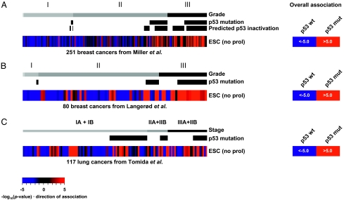
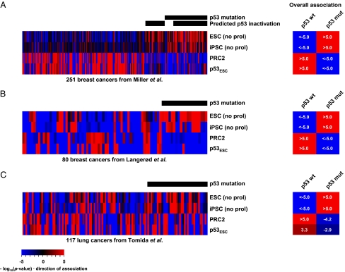
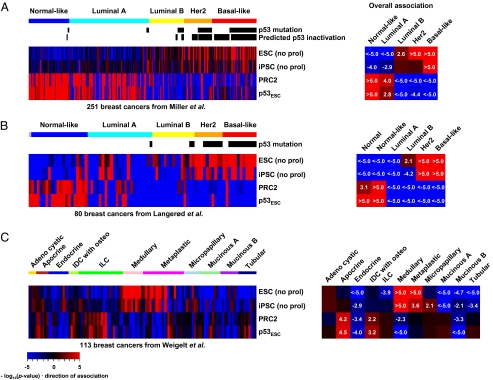
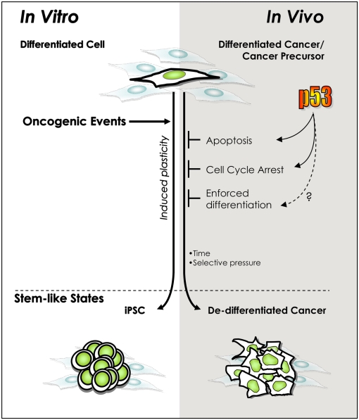
Breast cancer comprises a heterogeneous set of diseases distinguishable from one another by pathologic presentation and molecular signatures. However, each breast cancer subtype is also heterogeneous. Some of the heterogeneity may be attributable to genetic instability, but recent data emphasize that developmental plasticity may also contribute. The p53 tumor suppressor could constitute a nodal control point underlying both sources of heterogeneity because it is frequently inactivated during malignant progression and has recently been shown to function as a potent barrier preventing fully differentiated cells from reverting to pluripotent stem cells after expression of appropriate oncogenes. Using archival microarray datasets, we tested the hypothesis that a p53 mutation could allow cells within a tumor to acquire a stem cell-like state by looking for coordinate expression of stem cell identity genes. We show that breast and lung cancers with p53 mutations do exhibit stem cell-like transcriptional patterns. Such tumors were also depleted for differentiation genes regulated by the polycomb repressor complex 2. These data are consistent with a model in which loss of p53 function enables acquisition of stem cell properties, which are positively selected during tumor progression.


Conflict of interest statement

The authors declare no conflict of interest.

Figures

Fig. 1.
Fig. 1.
Association scores for the ESC signature in p53-sequenced cancers. (A) Analysis of 251 breast cancers from the Miller et al. dataset (18). Color saturation reflects both strength and direction of association (Materials and Methods); red indicates that genes in the signature are coordinately up-regulated in the sample and blue indicates down-regulation. Grade, p53 mutation status, and predicted p53 inactivation status are indicated. In addition, overall association (Right) was assessed using representative profiles generated by averaging the gene expression levels among p53 wild-type and p53 mutant tumors, respectively. Numerical values in A represent actual association scores defined as −log10(P value) × direction of association (Materials and Methods). (B) Association scores for the ESC signature in a second independent breast cancer dataset from Langerød et al. (19). (C) Association scores in the lung adenocarcinoma dataset from Tomida et al. (20), including information for p53 mutation status and tumor stage.
Fig. 2.
Fig. 2.
Additional metrics for the stem-like state correlate with p53 status. Association scores are shown for ESC, iPSC, PRC2, and p53ESC signatures in the (A) Miller et al. (18), (B) Langerød et al. (19), and (C) Tomida et al. datasets (20). Overall associations were determined as in Fig. 1.
Fig. 3.
Fig. 3.
Association scores for ESC, iPSC, PRC2, and p53ESC signatures in breast cancer subtypes. (A) Breast cancers were ordered according to the “intrinsic subtypes” from the Miller et al. dataset (18) and (B) the Langerød et al. dataset (19). (C) Association scores in the “11 special subtypes” of breast cancers from the Weigelt et al. dataset (25). IDC with osteo, invasive ductal carcinoma with osteoclastic giant cells; ILC, invasive lobular carcinoma. Overall associations were assessed using representative profiles generated by averaging the gene expression levels among tumors of each cancer subtype.
Fig. 4.
Fig. 4.
Model of parallel, p53-regulated, cellular de-differentiation processes in vitro and in vivo. The oncogenic lesions initiating and promoting tumorigenesis, like those inducing pluripotency in vitro, generally activate the tumor-suppressive p53 pathway. Consequently, inactivating p53 should permit survival and division under adverse conditions and time for the accumulation of an undefined number of epigenetic modifications to engender reprogramming. This model is similar to one proposed in response to reports that p53 inactivation impacts the efficiency of induced pluripotency in vitro (–13, 29). The present work provides evidence that a parallel role exists during tumorigenesis in vivo.

References

    1. Merlo LM, Pepper JW, Reid BJ, Maley CC. Cancer as an evolutionary and ecological process. Nat Rev Cancer. 2006;6:924–935. - PubMed
    1. Vogelstein B, Kinzler KW. The multistep nature of cancer. Trends Genet. 1993;9:138–141. - PubMed
    1. Stingl J, Caldas C. Molecular heterogeneity of breast carcinomas and the cancer stem cell hypothesis. Nat Rev Cancer. 2007;7:791–799. - PubMed
    1. Gupta PB, Chaffer CL, Weinberg RA. Cancer stem cells: Mirage or reality? Nat Med. 2009;15:1010–1012. - PubMed
    1. Takahashi K, Yamanaka S. Induction of pluripotent stem cells from mouse embryonic and adult fibroblast cultures by defined factors. Cell. 2006;126:663–676. - PubMed

Publication types

MeSH terms

Substances