

doi: 10.4103/0301-4738.73688.
Evaluation of a glaucoma patient
Affiliations
- PMID: 21150033
- PMCID: PMC3038503
- DOI: 10.4103/0301-4738.73688
Item in Clipboard
Evaluation of a glaucoma patient
Indian J Ophthalmol.
2011 Jan.
Abstract
The diagnosis of glaucoma is usually made clinically and requires a comprehensive eye examination, including slit lamp, applanation tonometry, gonioscopy and dilated stereoscopic evaluation of the optic disc and retina. Automated perimetry is obtained if glaucoma is suspected. This establishes the presence of functional damage and provides a baseline for follow-up. Imaging techniques are not essential for the diagnosis but may have a role to play in the follow-up. We recommend a comprehensive eye examination for every clinic patient with the objective of detecting all potentially sight-threatening diseases, including glaucoma.
Conflict of interest statement
Figures
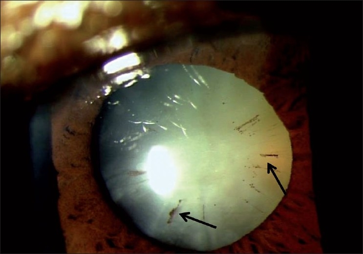
“Brown” stage of pseuduexfoliation (Arrow indicates “brown” stage of PXF)
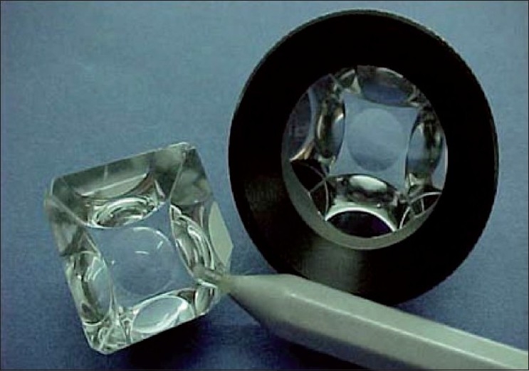
Indentation gonioscope
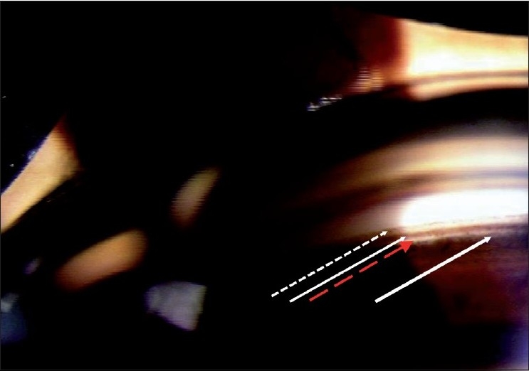
Normal angle anatomy (Broken arrow: Schwalbe’s line, White arrrow: Pigmented Trabecular Meshwork, Red arrow: Scleral Spur, Thick white arrow: Cilliary Body)
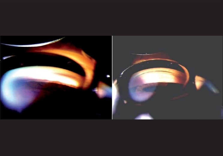
Gonioscopy showing angle with bright and dim illumination (Angle is open with bright illumination and the same angle is closed with appropriate testing conditons)
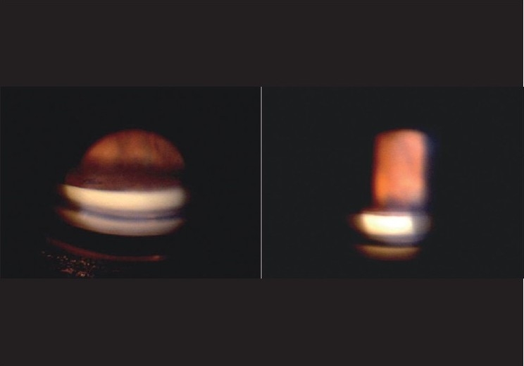
Gonioscopy showing “over the hill” angle (Gonioscopy showint no angle structure in straight ahead view. By tilting angle, “over the hill” view showing angle structures)
Gonioscopy showing peripheral anterior synechiae (Arrow showing peripheral anterior synechia)
Relation between optic disc size and cup [(a) Small disc and has a small cup. (b) Medium sized disc with a larger cup. (c) Large disc and a large cup]
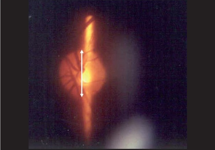
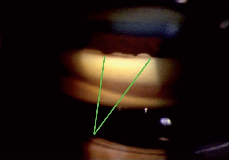
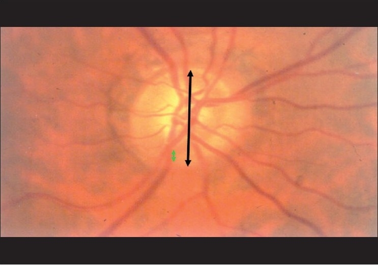
Estimation of optic disc size (Narrrow the vertical height of the slit beam to match the disc height)
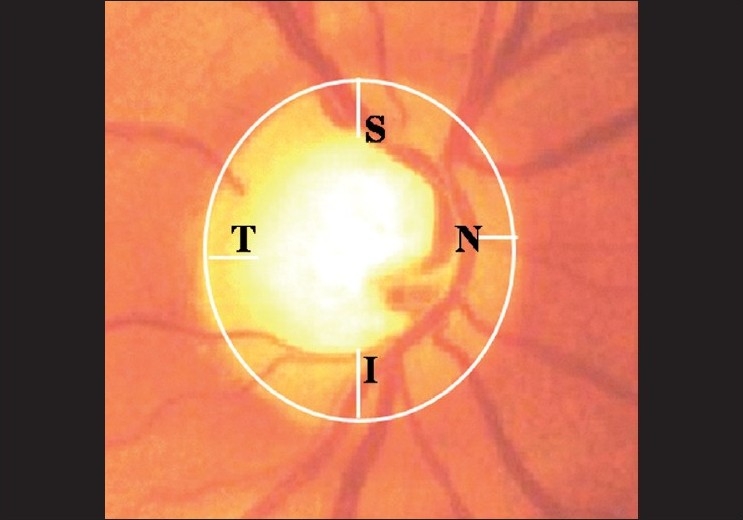
Neuro retinal rim: ISNT rule (Normally, inferior rim is thicker than superior rim, which in turn is thicker than nasal rim. Temporal rim is thinnest)
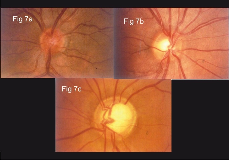
Optic disc showing early glaucoma (Loss of ISNT rule. Note that superior rim is thicker than inferior rim)

Optic disc with NOTCH
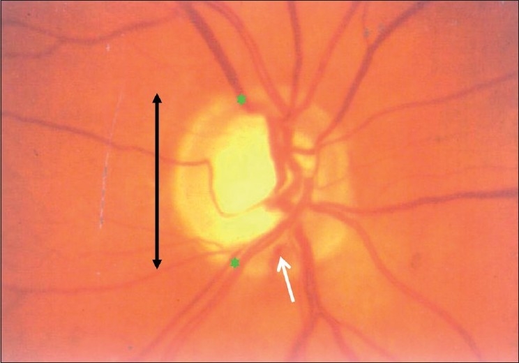
Optic disc showing disc hemorrhage (Rim to disc ratio <0.1:1 as seen here indicates glaucoma. Whith arrow indicates disc haemorrhage)
Red free optic disc photograph (Normal RNFL pattern: Bright dark bright pattern)
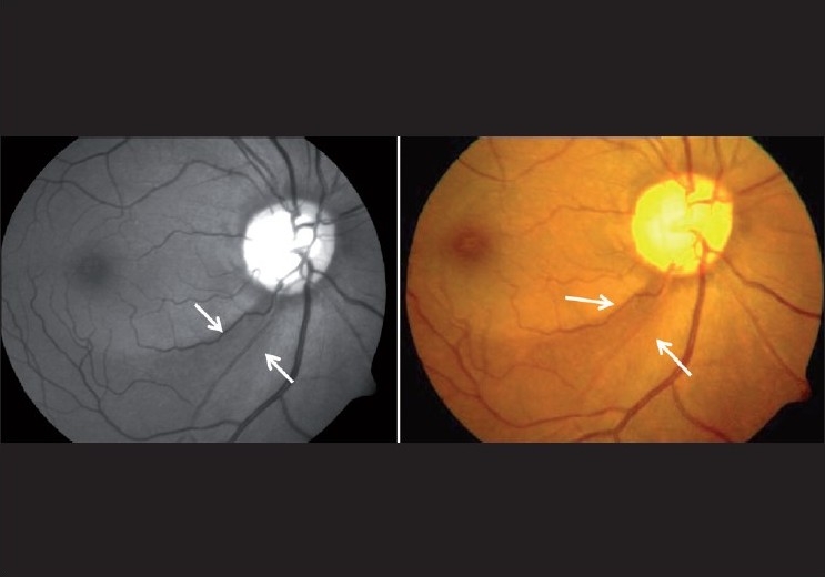
Optic photograph showing wedge-shaped defect (Arrow indiectes wedge shaped RNFL defects)
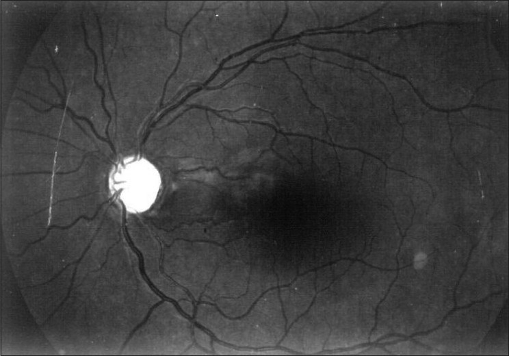
Red free photograph showing diffuse nerve fiber layer defects (Note that bright dark bright pattern is lost and it appears completely dark)
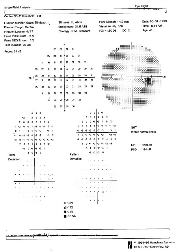
Normal visual field
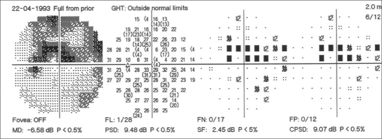
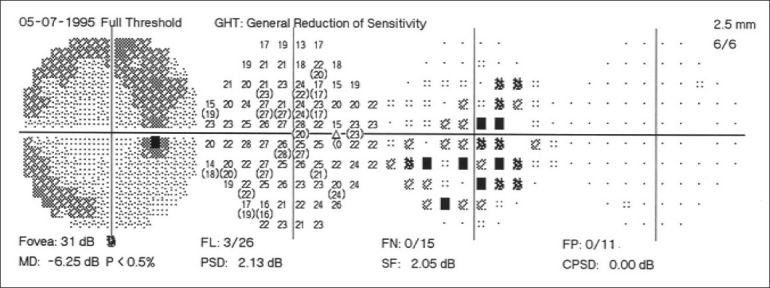
Visual field with an early glaucomatous defect
Visual field in patients with media opacity
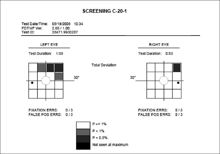
Glaucomatous visual field defect in the FDP 20 screening program (POAG patient with superior arcuate scotoma on WWP)
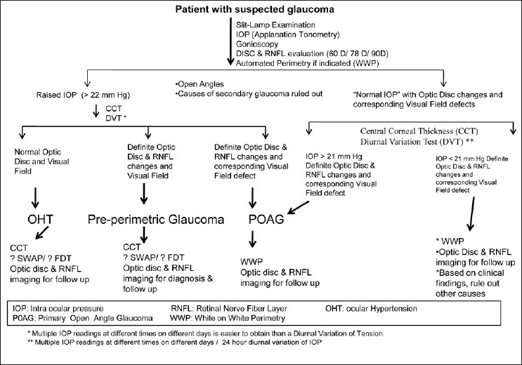
Evaluation of a glaucoma suspect (open angle glaucoma)
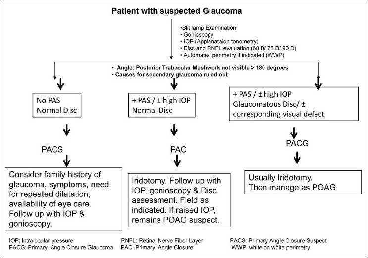
Evaluation of a glaucoma suspect (angle closure disease)
References
-
- Thomas R, Parikh R, Paul P, Muliyil J. Population-based screening versus case detection. Indian J Ophthalmol. 2002;50:233–7. - PubMed
-
- Grave E, Wienereb R, editors. Hague, Netherlands: Kurger Publication; 2004. Glaucoma Diagnosis: Structure and Function.
-
- San Francisco: CA: American Academy of Ophthalmology; 1996. American Academy of Ophthalmology. Primary Open-Angle Glaucoma. Preferred Practice Pattern. San Francisco.
-
- SEAGIG Asia Pacific Glaucoma Guidelines. 2nd ed. Available from: http://www.seagig.org[last cited on 2008-09]
-
- Healey P, Thomas R, editors. Oxford UK: Health Press ltd; 2009. The fast facts in glaucoma; pp. 1–128.
MeSH terms
LinkOut - more resources
Full Text Sources
Medical
