Review
. 2010 Nov-Dec;4(6):483-9.
doi: 10.4161/chan.4.6.12868. Epub 2010 Nov 1.

The neuronal splicing factor Nova controls alternative splicing in N-type and P-type CaV2 calcium channels

Affiliations
Review

The neuronal splicing factor Nova controls alternative splicing in N-type and P-type CaV2 calcium channels

Summer E Allen et al. Channels (Austin). 2010 Nov-Dec.

Abstract

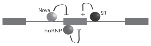
Many cellular processes are involved in optimizing protein function for specific neuronal tasks; here we focus on alternative pre-mRNA splicing. Alternative pre-mRNA splicing gives cells the capacity to modify and selectively re-balance their existing pool of transcripts in a coordinated way across multiple mRNAs, thereby effecting relatively rapid and relatively stable changes in protein activity. Here we report on and discuss the coordinated regulation of two sites of alternative splicing, e24a and e31a, in P-type CaV2.1 and N-type CaV2.2 channels. These two exons encode 4 and 2 amino acids, respectively, in the extracellular linker regions between transmembrane spanning segments S3 and S4 in domains III and IV of each CaV2 subunit. Recent genome-wide screens of splicing factor-RNA binding events by Darnell and colleagues show that Nova-2 promotes inclusion of e24a in CaV2.2 mRNAs in brain. We review these studies and show that a homologous e24a is present in theCaV2 .1 gene, Cacna1a, and that it is expressed in different regions of the nervous system. Nova-2 enhances inclusion of e24a but represses e31a inclusion in CaV2.1 and CaV2.2 mRNAs in brain. It is likely that coordinated alternative pre-mRNA splicing across related CaV2 genes by common splicing factors, allows neurons to orchestrate changes in synaptic protein function while maintaining a balanced and functioning system.


Figures

Figure 1
Figure 1
Nova acts as a splicing repressor or enhancer depending on its binding site relative to the target exon. Nova binds to YCAY motifs in introns upstream of e31a in CaV2 pre-mRNAs to repress its inclusion, whereas Nova binds to YCAY motifs in introns downstream of e24a in CaV2 pre-mRNAs to enhance its inclusion, during pre-mRNA splicing.
Figure 2
Figure 2
Nova binding motifs are located in the intron downstream of e24a of CaV2.2. The genomic sequence in this region of Cacna1b is shown, and the amino acids encoded by e24a indicated (SFMG). The Nova binding motifs (black squares) are shown below the genomic sequence and were mapped to the UCSC genome browser (July 2007 mouse assembly). Nova binding motifs are shown within 70 nucleotides and downstream of e24a in Cacna1b. The Nova binding site track was provided by SFmap. SFmap is a computational tool that uses an algorithm to predict splicing factor binding sites by identifying binding site motif clusters and evaluating the conservation of these clusters across species.
Figure 3
Figure 3
Alternatively spliced exons in the CaV2 genes. The locations of constitutive exons (black) and conserved alternative exons (colored) in mouse Cacna1a, Cacna1b and Cacna1e genes. Gene diagrams are adapted from the UCSC genome browser (Feb. 2006 mouse assembly). Alternative exons 37a and 37b are found in all three CaV2 channel genes. An alternative exon e18a is present in Cacna1b and Cacna1e, but a homologous exon has not been identified in Cacna1a. Alternative exons e24a and e31a are conserved in Cacna1a and Cacna1b, but equivalent exons have not been identified in Cacna1e.
Figure 4
Figure 4
Alternative exons e31a of CaV2.1 and CaV2.2 are repressed in brain by Nova-2. The approximate locations of e31a of CaV2.1 (NP) and CaV2.2 (ET) are mapped on a schematic of CaV2 channels that shows transmembrane and intracellular domains (A and B). RT-PCR products amplified from mouse brain using primers that flank e31a are shown separated in 8% denaturing polyacrylamide gels. Predicted sizes of PCR products are 100 (+31a) and 94 (Δ31a) nts for CaV2.1 (A) and 106 (+31a) and 100 (Δ31a) nts for CaV2.2 (B). RT-PCR amplification was carried out using brain lacking cerebellum (Brain), cerebellum (CB) and dorsal root ganglia (DRG) from wild-type (WT) and Nova-2 knockout mice (KO). Percentages of cDNAs containing e31a of the total amplified cDNA for each reaction are shown below each lane. (A) 30% of cDNAs amplified from wild-type brain contain e31a compared to 80% in Nova-2-/- brain. In cerebellum, 17% of cDNAs amplified from wild-type tissue contain e31a compared to 46% in Nova-2-/- tissue. cDNA amplified from DRG contains a high percentage of e31a-containing sequence similarly in wild-type and Nova-2 knockout tissue (82–87%). (B) cDNAs amplified from wild-type brain lacked e31a compared to 44% representation in cDNAs from Nova-2-/- brain. In cerebellum, 13% of cDNAs amplified from wild-type tissue contain e31a compared to 32% in Nova-2-/- tissue. cDNA amplified from DRG contains a high percentage of e31a-containing sequence (83–84%) similarly in wild-type and Nova-2 knockout tissue. A schematic shown below the gels illustrates how Nova acts to repress e31a inclusion via a binding site in the upstream intron in brain and cerebellum, but not in DRG.
Figure 5
Figure 5
Alternative exons e24a of CaV2.1 and CaV2.2 are enhanced in brain by Nova-2. (A) Location and sequence, SSTR, of newly identified e24a (red) in the mouse Cacna1a gene. Constitutive exons e24 and e25 are shown in gray flanking the alternatively spliced e24a (red). Below, the degree of sequence conservation among vertebrate Cacna1a genes is represented. Image is adapted from UCSC genome browser, Feb 2006 mouse assembly. (B) RT-PCR products amplified using primers located in e24 and e25 from RNA isolated from the following regions of adult mouse brain: cortex (CTX), hippocampus (HC), hypothalamus (HP), midbrain (MB), olfactory bulb (OB) and cerebellum (CB). Predicted sizes of products are 152 nts (+24a) and 140 nts (Δ24a). PCR products were separated on 8% denaturing polyacrylamide gels. (C) RT-PCR products amplified from mouse brain using primers that flank e24a are shown separated on a 3% agarose (DRG) or 8% denaturing polyacrylamide gels. Predicted sizes of PCR products are 152 (+24a) and 140 (Δ24a) nts. RT-PCR amplification was carried out using brain lacking cerebellum (Brain), cerebellum (CB) and dorsal root ganglia (DRG) from wild-type (WT) and Nova-2 knockout mice (KO). Percentages of cDNAs containing e24a of the total amplified cDNA pool are shown below each lane. (A) 13% of cDNAs amplified from wild-type brain contain e24a but this isoform was not detectable in Nova-2-/- brain samples. In cerebellum, 23% of cDNAs amplified from wild-type tissue contain e24a compared to 12% in Nova-2-/- tissue. cDNA amplified from DRG contains a very low percentage of e24a-containing sequence similarly in wild-type and Nova-2 knockout tissue (4–5%). A schematic shown below the gels illustrates how Nova acts to enhance e24a inclusion via a binding site in the downstream intron in brain and cerebellum, but not in DRG.
Figure 6
Figure 6
Cellular control of alternative splicing likely involves the net contribution of more than one splicing factor. Schematic shows hypothetical, combinatorial control of an alternatively spliced exon by Nova binding downstream and acting as a repressor, by another serine rich (SR) splicing factor binding downstream and acting as an enhancer, and by a third splicing factor of the hnRNP family binding to the target exon and acting as a repressor of exon inclusion. The relative importance and contribution of each splicing factor depends on several factors, including their expression levels. Thus, tissue specific splicing can arise from the tissue-specific expression profiles of splicing factors.

References

    1. Le Hir H, Seraphin B. EJCs at the heart of translational control. Cell. 2008;133:213–216. - PubMed
    1. Isken O, Maquat LE. Quality control of eukaryotic mRNA: safeguarding cells from abnormal mRNA function. Genes Dev. 2007;21:1833–1856. - PubMed
    1. McGlincy NJ, Smith CW. Alternative splicing resulting in nonsense-mediated mRNA decay: what is the meaning of nonsense? Trends Biochem Sci. 2008;33:385–393. - PubMed
    1. Boutz PL, Stoilov P, Li Q, Lin CH, Chawla G, Ostrow K, et al. A post-transcriptional regulatory switch in polypyrimidine tract-binding proteins reprograms alternative splicing in developing neurons. Genes Dev. 2007;21:1636–1652. - PMC - PubMed
    1. Makeyev EV, Zhang J, Carrasco Ma, Maniatis T. The MicroRNA miR-124 promotes neuronal differentiation by triggering brain-specific alternative pre-mRNA splicing. Mol Cell. 2007;27:435–448. - PMC - PubMed

Publication types

MeSH terms

LinkOut - more resources