. 2010 Dec 15;9(24):4908-21.
doi: 10.4161/cc.9.24.14199. Epub 2010 Dec 15.

Proteomic profiling of Myc-associated proteins

Affiliations

Proteomic profiling of Myc-associated proteins

Pooja Agrawal et al. Cell Cycle. .

Abstract

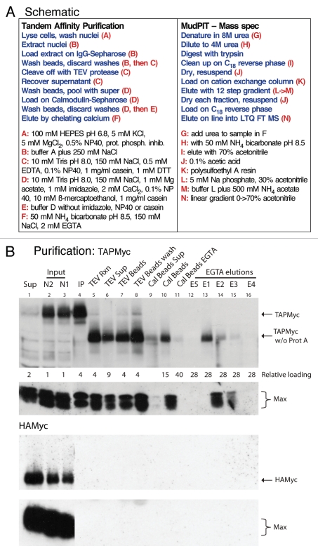
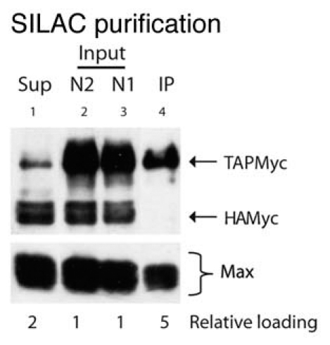
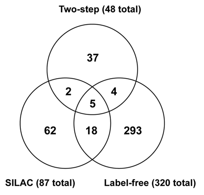
Mammalian c-Myc is a member of a small family of three closely related transcription factors. The Myc family of proto-oncogenes are among the most potent activators of tumorigenesis, and are frequently overexpressed in diverse cancers. c-Myc has an unusually broad array of regulatory functions, which include, in addition to roles in the cell cycle and apoptosis, effects on a variety of metabolic functions, cell differentiation, senescence, and stem cell maintenance. A significant number of c-Myc interacting proteins have already been defined, but it is widely believed that the c-Myc interactome is vastly larger than currently documented. In addition to interactions with components of the transcription machinery, transcription independent nuclear interactions with the DNA replication and RNA processing pathways have been reported. Cytoplasmic roles of c-Myc have also been recently substantiated. Recent advances in proteomics have opened new possibilities for the isolation of protein complexes under native conditions and confidently identifying the components using ultrasensitive, high mass accuracy and high resolution mass spectrometry techniques. In this communication we report a new tandem affinity purification (TAP) c-Myc interaction screen that employed new cell lines with near-physiological levels of c-Myc expression with multi-dimensional protein identification techniques (MudPIT) for the detection and quantification of proteins. Both label-free and the recently developed stable isotope labeling with amino acids in cell culture (SILAC) methodologies were used. Combined data from multiple biological replicates provided a dataset of 418 non-redundant proteins, 389 of which are putative novel interactors. This new information should significantly advance our understanding of this interesting and important master regulator.


Figures

Figure 1
Figure 1
Size exclusion chromatography of nuclear TAPMyc complexes. (A) Schematic representation of the TAPMyc protein. Structure of the TAP-tag: Prot A, protein A; TEV, TEV protease cleavage site; CBP, calmodulin binding protein. c-Myc contains a N-terminal transactivation domain and a C-terminal DNA binding domain. The conserved regions of c-Myc known as Myc boxes I through IV are shown a MBI, MBII, MBIII and MBIV. The C-terminal domain is composed of a basic region, b, a helix-loop-helix region, HLH, and a leucine zipper, LZ. In the MycTAP protein (not shown) the order of domains was reversed to be (N-to-C terminus): Myc(MBI-IV-b-HLH-LZ)-Cal BP-TEV-Prot A. (B) Size exclusion chromatography of TAPMyc-and MycTAP-containing nuclear exacts prepared using the Evan and Hancock method on a Superdex 200 column. Fractions were resolved by SDS-PAGE and immunoblotted for the protein A epitope of the TAP-tag. Lower portions of the same gel were probed for Max. Elution of molecular weight markers is shown above.
Figure 2
Figure 2
Tandem affinity purification of TAPMyc complexes. (A) For single-step purifications, after washing IgG beads with buffer B, beads were washed with buffer B without protease and phosphatase inhibitors, casein and NP40, and bound proteins were eluted with 100 mM glycine pH 2.5. The MudPIT/MS steps were the same after all purifications. (B) Purifications of nuclear extracts from TAPMyc (upper parts) and HAMyc (lower parts) expressing cells were monitored by immunoblotting for both c-Myc and Max at each purification step. The relevant proteins are identified in the right margin. Note the faster migration of the TAPMyc protein after removal of the protein A domain with TEV protease (TAPMyc w/o Prot A). Lane 1 (Sup), nuclear extract after pulldown with IgG beads; lanes 2 and 3 (N1 and N2), nuclei were extracted twice and the fractions were pooled as the starting material (Input) for the purification; lane 4 (IP), after pulldown the IgG beads were washed and an aliquot was boiled in sample buffer to remove bound Myc complexes (note efficient pulldown of Max, and lack of pulldown of either HAMyc or Max in the lower parts); lane 5 (TEV Rxn) IgG beads were incubated with TEV protease and an aliquot of the reaction was treated with sample buffer (note the almost complete cleavage of the TAPMyc protein); lane 6 (TEV Sup) beads were removed and the supernatant was assayed for released Myc complexes; lane 7 (TEV Beads) complexes remaining bound to the beads were released with sample buffer (note strong retention of complexes on beads in spite of efficient TEV cleavage); lane 8 (TEV beads wash) TEV beads were washed extensively with CB buffer and complexes remaining on beads were scored by release with sample buffer (note continuing strong retention of complexes on beads); lane 9 (Cal Beads Sup) the TEV-released complexes were loaded onto Calmodulin beads, the beads were spun down, and complexes remaining in the supernatant were assayed (note reasonable capture by Calmodulin beads); lane 10 (Cal beads) pulled-down complexes were determined by boiling an aliquot of the beads with sample buffer; lane 11 (Cal Beads EGTA) Cal Beads after elution with EGTA were treated as before (lane 10; note efficient release of complexes with EGTA); lanes 12–16 are individual fractions of the EGTA elutions (E1 through E5, in that order), note that most of the complexes eluted in fractions 1 and 2. Loading of lanes is indicated below the top part, for example, lane 16 contains the equivalent of 28-times the amount of starting material shown in lane 3. The lanes in the HAMyc parts are loaded in the same order and same proportions.
Figure 3
Figure 3
Characterization of TAPMyc purified complexes. (A) The Calmodulin Sepharose eluate was subjected to a series of spins: spin 1, 25,000 g, 20 min, spin 2, 50,000 g, 20 min, spin 3, 75,000 g, 20 min., spin 4, 100,000 g, 20 min. The pellet of each spin was recovered (P1 through P4) and the supernatant was carried over into the next spin. Supernatants: final, 100,000 g; start, input material. Samples were analyzed by immunoblotting; the relevant proteins are identified in the right margin. rMyc; recombinant c-Myc used as a loading control. (B) The Calmodulin Sepharose eluate was concentrated by centrifugal ultrafiltration (Microcon, Millipore) and subjected to size exclusion chromatography on a Superdex 200 column. Fractions were resolved by SDS-PAGE and immunoblotted. Because the TAPMyc protein at this stage of purification had the protein A epitope cleaved off, we had to rely on c-Myc antibodies to detect it. Although we could clearly detect TAPMyc in the input, because of the limited quantities this starting material and the relatively low affinity of available c-Myc antibodies, we were not able to cleanly detect TAPMyc in the column fractions. Max protein was however clearly visible. Elution of molecular weight markers is shown above.
Figure 4
Figure 4
SDS-PAGE and silver stain analysis of purified TAPMyc complexes. The Calmodulin Sepharose eluate was concentrated by centrifugal ultrafiltration (Microcon, Millipore) and 3 µg protein was run on a 3–10% polyacrylamide gel followed by silver staining. The c-Myc band is indicated in the left margin, as are additional bands that appeared enriched in the TAPMyc sample (arrows). IgG heavy and light chains are indicated in the right margin.
Figure 5
Figure 5
Single-step purification of TAPMyc complexes. TAPMyc and HAMyc cells, grown in light and heavy media respectively, were pooled in equal proportions and subjected to the Evan and Hancock nuclear extraction. Samples were analyzed by immunoblotting; the relevant proteins are identified in the right margin. Lane 1 (Sup), nuclear extract after pulldown with IgG beads; lanes 2 and 3 (N1 and N2), nuclei were extracted twice and the fractions were pooled as the starting material (Input) for the purification; lane 4 (IP), after pulldown the IgG beads were washed and an aliquot was boiled in sample buffer to remove bound Myc complexes (note efficient pulldown of Max, and lack of pulldown of HAMyc). The numbers shown below the parts indicate the relative loading of the individual lanes in comparison to the input.
Figure 6
Figure 6
Venn diagram of the relationships between the two-step, label-free and SILAC series of experiments. The overlap between the two-step and label-free datasets is 9 proteins (19% of two-step), between the two-step and SILAC datasets 7 proteins (15% of two-step), and between the SILAC and label-free datasets 23 proteins (27% of SILAC). The two-step dataset shares 11 proteins with the other two datasets (23%), the label-free dataset shares 27 proteins with the other two datasets (8%) and the SILAC dataset shares 25 proteins with the other two datasets (29%). Five proteins were found in all datasets (MAX, ERBB2IP, PRKCDBP, TUBA4A, UBC), and 24 additional proteins were present in two out of three datasets (ACTC1, ADNP, CBFB, CEP170, CKAP4, DNAJB12, EP400, ETV3, FLNA, FN1, HSPB1, LGALS1, LMNA, PSMD2, PTRF, QPCTL, RAB11FIP5, RIMS2, RPS27L, RUNX1, SERPINH1, SMTN, TXN, XYLT1). Proteins not found in the HPRD or Koch et al. datasets are underlined.

Comment in

References

    1. Meyer N, Penn LZ. Reflecting on 25 years with MYC. Nat Rev Cancer. 2008;8:976–990. - PubMed
    1. Albihn A, Johnsen JI, Henriksson MA. MYC in oncogenesis and as a target for cancer therapies. Adv Cancer Res. 2010;107:163–224. - PubMed
    1. Dai MS, Jin Y, Gallegos JR, Lu H. Balance of Yin and Yang: ubiquitylation-mediated regulation of p53 and c-Myc. Neoplasia. 2006;8:630–644. - PMC - PubMed
    1. Ponzielli R, Katz S, Barsyte-Lovejoy D, Penn LZ. Cancer therapeutics: Targeting the dark side of Myc. Eur J Cancer. 2005;41:2485–2501. - PubMed
    1. Cole MD, Cowling VH. Transcription-independent functions of MYC: Regulation of translation and DNA replication. Nat Rev Mol Cell Biol. 2008;9:810–815. - PMC - PubMed

Publication types

MeSH terms