Review
. 2011 Jan;11(1):68-75.
doi: 10.1038/nrc2950. Epub 2010 Dec 2.

What is the malignant nature of human ductal carcinoma in situ?

Affiliations
Review

What is the malignant nature of human ductal carcinoma in situ?

Virginia Espina et al. Nat Rev Cancer. 2011 Jan.

Abstract

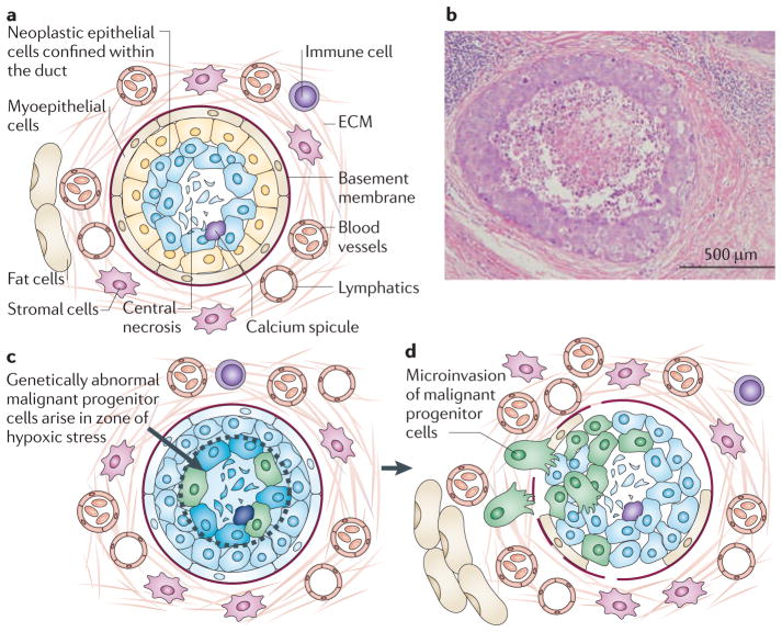
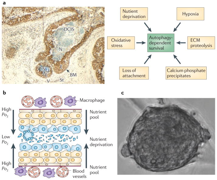
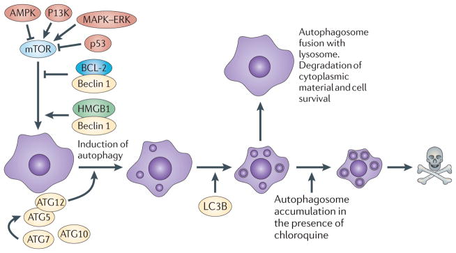
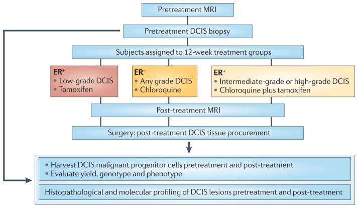
Invasive, genetically abnormal carcinoma progenitor cells have been propagated from human and mouse breast ductal carcinoma in situ (DCIS) lesions, providing new insights into breast cancer progression. The survival of DCIS cells in the hypoxic, nutrient-deprived intraductal niche could promote genetic instability and the derepression of the invasive phenotype. Understanding potential survival mechanisms, such as autophagy, that might be functioning in DCIS lesions provides strategies for arresting invasion at the pre-malignant stage. A new, open trial of neoadjuvant therapy for patients with DCIS constitutes a model for testing investigational agents that target malignant progenitor cells in the intraductal niche.


Conflict of interest statement

Competing interests statement

The authors declare no competing financial interests.

Figures

Figure 1
Figure 1. The stressful microenvironment of the intraductal niche may promote genetic instability
a | This figure shows the microecology of ductal carcinoma in situ (DCIS) lesions in the breast. DCIS neoplastic cells proliferate within the duct, which is bound by the basement membrane and a rim of myoepithelial cells on the lumen side of the basement membrane. The normal duct is composed of a single epithelial layer. By contrast, the DCIS lesion contains multiple layers of cells that accumulate inwards into the lumen. Outside the basement membrane the breast stroma contains the extracellular matrix (ECM), lymphatics, blood vessels, stromal cells, immune cells and fat cells. All of these components participate in the carcinogenic process by promoting or suppressing malignant progenitor cells that could arise within the mass of cells accumulating in the duct. b | Haematoxylin and Eosin stained section (original magnification x10) of a comedo-DCIS lesion, exhibiting central necrosis, stroma and lymphocytes. c | Necrosis occurs in the centre of the duct because these cells are the furthest radial distance from the oxygen and nutrients that diffuse from the vessels outside the duct. Proliferating cells within the intraductal microenvironment are under high stress because they are nutrient deprived, hypoxic, crowded and undergoing oxidative stress. d | Mammary pluripotent stem cells must adapt to survive in the high-stress microenvironment. Adaptation promotes the suppression of apoptosis in the face of genetic instability and could lead to the generation of genetically abnormal malignant progenitor cells before the onset of invasion. Invasion is associated with the loss of myoepithelial cells, periductal angiogenesis, fragmentation of the basement membrane and chemotaxis radially outwards.
Figure 2
Figure 2. Autophagy and cell survival in DCIS lesions
a | Human comedo-ductal carcinoma in situ (DCIS) lesion (original magnification x10) stained with antibodies to Beclin 1 shows upregulation of Beclin 1 (brown staining) in the viable rim of intraductal cells within the hypoxic ductal niche. Multiple types of stress that impinge on the DCIS cells directly contribute to the activation of autophagy (arrows; right side of figure). b | As abnormal DCIS cells accumulate within the duct many are pushed further from the source of oxygen and nutrients outside the duct. This sets up a gradient of stress that could be a ‘breeding ground’ for the mutational and cytogenetic abnormalities that drive breast cancer. Understanding how genetically abnormal DCIS cells arise and survive within the high-stress microenvironment provides targets for chemoprevention. c | Example of a human malignant precursor spheroid that can be propagated from fresh DCIS tissue. BM, basement membrane; E, epithelial cells; ECM, extracellular matrix; N, necrosis; St, stroma; V, vessels.
Figure 3
Figure 3. Upstream pathways that intersect with the autophagic pathway
Autophagy is activated and regulated by various molecules that are known to be involved in oncogenesis, such as AMP-activated protein kinase (AMPK), PI3K and p53. Autophagy is a catabolic process involving the degradation of the components of a cell by its own lysosomal machinery. Autophagy is a major mechanism by which a starving cell reallocates nutrients from unnecessary processes to more essential processes. The best-studied mechanism of autophagy involves the nucleation of a double-membrane vesicle around a cellular constituent. The resultant vesicle then fuses with a lysosome that contains acid proteases that degrade the contents, ultimately generating ATP for the cell. Chloroquine treatment blocks autophagy by interfering with the fusion of the autophagosome vesicle with the lysosome and by altering the acidic internal pH of the lysosome. ATG12, autophagy 12; HMGB1, high mobility group protein B1; LC3B, microtubule-associated protein 1 light chain 3β.
Figure 4
Figure 4. A neoadjuvant therapy trial for DCIS
The Preventing Invasive Neoplasia with Chloroquine (PINC) trial (NCT01023477; see Further information) examines the safety and effectiveness of chloroquine administration to patients with low-grade, intermediate-grade or high-grade ductal carcinoma in situ (DCIS). Patients with high-grade DCIS whose lesion is oestrogen receptor-positive (ER+) will receive standard of care tamoxifen plus chloroquine. Patients who are ER will receive chloroquine alone. Patients with ER+, low-grade lesions will receive tamoxifen only. Magnetic resonance imaging (MRI) will be carried out on each patient before enrolment and just before the standard-of-care surgical therapy, which will be mastectomy or lumpectomy depending on the size and confluence of the primary DCIS lesion. Effectiveness in this DCIS trial design will be uniquely measured directly at the molecular level in the DCIS tissue before and after treatment. The genotype, phenotype and proteome of the harvested malignant progenitor cells from the DCIS lesions, and the molecular histology of the DCIS lesion, will be compared with MRI images before and after a 3-month course of therapy.

Comment in

References

    1. Allegra C, et al. NIH state-of-the-science conference statement: diagnosis and management of ductal carcinoma in situ. NIH Consens State Sci Statements. 2009;26:1–27. - PubMed
    1. Castro NP, et al. Evidence that molecular changes in cells occur before morphological alterations during the progression of breast ductal carcinoma. Breast Cancer Res. 2008;10:R87. - PMC - PubMed
    1. Damonte P, et al. Mammary carcinoma behavior is programmed in the precancer stem cell. Breast Cancer Res. 2008;10:R50. - PMC - PubMed
    1. Espina V, et al. Malignant precursor cells pre-exist in human breast DCIS and require autophagy for survival. PLoS ONE. 2010;5:e10240. - PMC - PubMed
    1. Ma XJ, Dahiya S, Richardson E, Erlander M, Sgroi DC. Gene expression profiling of the tumor microenvironment during breast cancer progression. Breast Cancer Res. 2009;11:R7. - PMC - PubMed

Publication types