. 2011 Jun;18(6):948-58.
doi: 10.1038/cdd.2010.162. Epub 2010 Dec 10.

p75 neurotrophin receptor mediates apoptosis in transit-amplifying cells and its overexpression restores cell death in psoriatic keratinocytes

Affiliations

p75 neurotrophin receptor mediates apoptosis in transit-amplifying cells and its overexpression restores cell death in psoriatic keratinocytes

F Truzzi et al. Cell Death Differ. 2011 Jun.

Abstract

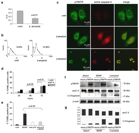
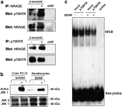
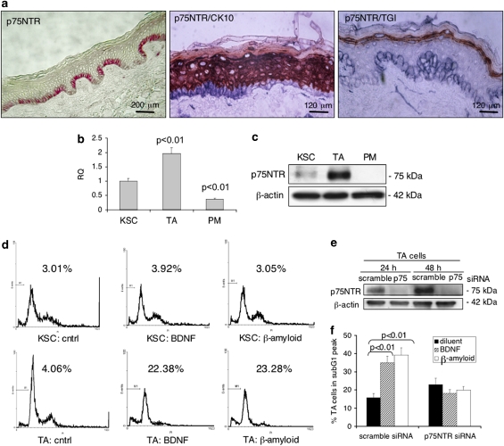
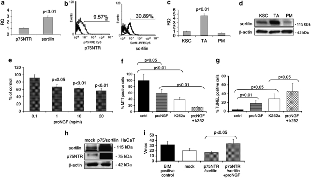
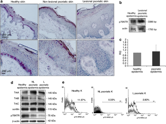
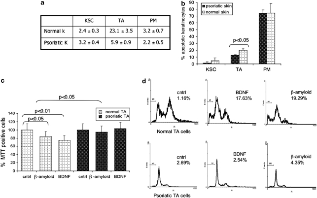
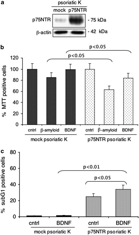
p75 neurotrophin receptor (p75NTR) belongs to the TNF-receptor superfamily and signals apoptosis in many cell settings. In human epidermis, p75NTR is mostly confined to the transit-amplifying (TA) sub-population of basal keratinocytes. Brain-derived neurotrophic factor (BDNF) or neurotrophin-4 (NT-4), which signals through p75NTR, induces keratinocyte apoptosis, whereas β-amyloid, a ligand for p75NTR, triggers caspase-3 activation to a greater extent in p75NTR transfected cells. Moreover, p75NTR co-immunoprecipitates with NRAGE, induces the phosphorylation of c-Jun N-terminal kinase (JNK) and reduces nuclear factor kappa B (NF-κB) DNA-binding activity. p75NTR also mediates pro-NGF-induced keratinocyte apoptosis through its co-receptor sortilin. Furthermore, BDNF or β-amyloid cause cell death in TA, but not in keratinocyte stem cells (KSCs) or in p75NTR silenced TA cells. p75NTR is absent in lesional psoriatic skin and p75NTR levels are significantly lower in psoriatic than in normal TA keratinocytes. The rate of apoptosis in psoriatic TA cells is significantly lower than in normal TA cells. BDNF or β-amyloid fail to induce apoptosis in psoriatic TA cells, and p75NTR retroviral infection restores BDNF- or β-amyloid-induced apoptosis in psoriatic keratinocytes. These results demonstrate that p75NTR has a pro-apoptotic role in keratinocytes and is involved in the maintenance of epidermal homeostasis.


Figures

Figure 1
Figure 1
p75NTR modulates apoptosis in normal human keratinocytes in vitro. (a) Confluent keratinocyte cultures, seeded in a 96-well tissue culture plate, were treated with 40 μM β-amyloid or diluent alone. MTT assay was performed after 24 h. Results are expressed as the mean±S.D. of three different experiments. Student's t-test was used for comparison of the means. (b) Confluent cells were treated as described before, trypsinized at 24 h and stained with PI. Cells in subG1 peak were analyzed by flow cytometry. (c) Keratinocytes cultures, seeded on chamber slides, were provided with 40 μM β-amyloid or diluent for 24 h. Cells were fixed, double stained in situ with anti-p75NTR and anti-active caspase-3 antibodies, and analyzed by a Confocal Scanning Laser Microscopy. (d) Confluent keratinocyte cultures were treated with BDNF or NT-4 (100 ng/ml). At 24, 48 and 72 h cells were fixed and stained in situ with TUNEL. Positive cells were counted as described in Materials and Methods section. (e) Confluent keratinocytes were treated individually or in combination with 100 ng/ml BDNF and 50 μg per ml of mouse anti-p75NTR neutralizing antibody. After 48 h, cells were stained in situ with TUNEL and positive cells were counted as described in Materials and Methods section. (f) Keratinocytes were transiently infected with p75NTR-LNSN packaging cells or LNSN packaging cells as mock control. Both mock and transfected cells were treated with BDNF (100 ng/ml) or β-amyloid (40 μM). At 48 h after treatment, protein extracts from keratinocytes were analyzed by western blotting for p75NTR and caspase-3 expression. β-actin was used as internal control. (g) Relative intensity of bands on radiograms was quantified by laser scanner densitometry. Values of pro-caspase-3 and caspase-3 fragment are expressed as fold variations compared with mock-diluent keratinocytes
Figure 2
Figure 2
p75NTR induces apoptosis via JNK and NF-κB. (a) Keratinocytes were treated for 24 h with β-amyloid (40 μM) or diluent. Protein extracts were immunoprecipitated with NRAGE serum and immunoblotted with p75NTR antibody and with NRAGE serum as control. Cell lysates were also immunoprecipitated with p75NTR antibody, immunoblotted with NRAGE serum and with p75NTR antibody as control. (b) Confluent keratinocytes were treated for 5 min with BDNF (100 ng/ml) and protein extracts were analyzed by western blotting using anti-active JNK1 polyclonal antibody. PC12 cells treated with 0.5 M sorbitol for 5 min were used as positive control. (c) Keratinocytes were infected with p75NTR and treated with with 100 ng/ml BDNF or diluent alone for 12 h. Nuclear protein extracts were analyzed by Gel mobility shift assay for NF-κB DNA-binding activity
Figure 3
Figure 3
p75NTR-induced apoptosis in TA cells. (a) Cryosections of normal skin were acetone-fixed and stained with anti-p75NTR antibody. Fast red was used as cromogen for p75NTR. Cryosections were double stained with anti-p75NTR and anti-keratin 10 (CK10) antibodies or with anti-p75NTR and transglutaminase type I (TgaseI) antibodies. Fast blue was used as cromogen for p75NTR, whereas carbazol for CK10 and TGaseI. (b) Normal human keratinocytes were obtained from neonatal foreskin and separated into three sub-populations on type IV collagen. A real-time PCR was performed on RNA extracts of the three populations by using primers for p75NTR, as described in Materials and Methods section. KSC were used as calibrator. Student's t-test was performed between samples and calibrator. (c) Protein extracts from the three populations were separated on 7% polyacrylamide gels and transferred onto nitrocellulose membrane. Membranes were immunoblotted with anti-p75NTR antibody. β-actin was used as internal control. (d) KSC and TA cells were treated with either BDNF or β-amyloid, stained with PI, and analyzed by flow cytometry. (e) TA cells were transiently transfected with 50 nM of p75NTR siRNA, as shown by western blotting. (f) At 48 h after transfection, both mock and transfected cells were treated with BDNF (100 ng/ml) or β-amyloid (40 μM). SubG1 peak analysis was performed after 24 h
Figure 4
Figure 4
Pro-NGF mediates apoptosis in human keratinocytes through p75NTR and sortilin. (a) Normal human keratinocytes were obtained from neonatal foreskin. A real-time PCR was performed on RNA extracts by using primers for p75NTR and sortilin, as described in Materials and Methods section. p75NTR expression levels were used as calibrator. (b) Keratinocyte cultures were trypsinized and stained with mouse monoclonal anti-p75NTR and polyclonal anti-sortilin antibody. Cells were analyzed by flow cytometry. (c) Normal human keratinocytes were separated into three sub-populations on type IV collagen. Real-time PCR was performed on RNA extracts of the three populations by using primers for sortilin, as described in Materials and Methods section. KSCs were used as calibrator. (d) Protein extracts from the three populations were separated on 7% polyacrylamide gels and transferred onto nitrocellulose membrane. Membranes were immunoblotted with anti-sortilin antibody. (e) Keratinocyte cultures, seeded in a 96-well tissue culture plate, were treated with 0.1, 1, 10, 20 ng/ml of pro-NGF. MTT assay was performed after 72 h. (f) Keratinocyte confluent cultures, seeded in a 96-wells tissue culture plate, were treated with 10 ng/ml of pro-NGF or 200 nM k252a alone, or in combination. MTT assay was performed after 72 h. (g) Confluent keratinocytes, seeded in a tissue culture slide-flask, were treated as described above. At 72 h, cells were fixed and stained in situ with TUNEL. Positive cells were counted as described in Materials and Methods section. (h) HaCaT cells were transfected with 2 μg of pcDNA3-BimEL (positive control) in combination with 2 μg of pcDNA3-p75NTR and pcDNA3-sortilin. Transfection was controlled by western blotting using anti-sortilin and anti-p75NTR antibody. (i) After 24 h, cells were treated with 10 ng/ml of pro-NGF for 24 h. Thereafter, HaCaT cells were lysed and caspase activity was assessed using DEVD-AFC substrate. Data represent the mean±S.E. of triplicate determinations
Figure 5
Figure 5
NT-receptor expression in psoriasis. (a) Cryosections of healthy, non-lesional and lesional psoriatic skin were acetone-fixed and stained with anti-p75NTR antibody. Fast red was used as cromogen. (b) mRNA extracts from healthy and psoriatic epidermis were analyzed by northern blot. DIG-labeled probes specific for p75NTR or actin were used. (c) Real-Time PCR was performed on healthy and psoriatic epidermal mRNA, as described in Materials and Methods section. Healthy mRNA was used as calibrator. (d) Protein extracts from healthy, lesional and non-lesional psoriatic epidermis were separated on 7% polyacrylamide gels and transferred onto nitrocellulose membrane. Membranes were immunoblotted with anti-p75NTR, anti-sortilin, anti-TrkA, anti-TrkB, anti-TrkC and β-actin antibodies. (e) Keratinocyte extracts from healthy, lesional and non-lesional psoriatic skin were stained with mouse monoclonal anti-p75NTR antibody. Cells, obtained from three different patients, were analyzed by flow cytometry
Figure 6
Figure 6
Reduced levels of p75NTR in TA cells are responsible for resistance to apoptosis in psoriatic keratinocytes. (a) Normal and psoriatic human keratinocytes were obtained from skin biopsies and separated into three sub-populations on type IV collagen. Keratinocytes were trypsinized and stained with mouse monoclonal anti-p75NTR and analyzed by flow cytometry. (b) After 48 h, both normal and psoriatic KSC, TA, and PM cells were stained in situ with PI and analyzed by Confocal Scanning Laser Microscopy. Data represent the mean±S.E. of triplicate determinations. (c) Keratinocyte cultures obtained from normal and psoriatic biopsies were treated with 40 μM β-amyloid or 100 ng/ml BDNF. MTT assay was performed after 48 h. (d) Keratinocytes were treated as shown in panel (c) and subG1 peak was evaluated by flow cytometry. Data represent the mean±S.E. from three independent experiments
Figure 7
Figure 7
p75NTR infection restores apoptosis susceptibility in psoriatic keratinocytes. (a) Psoriatic keratinocytes were double infected with mock or p75NTR retroviral vector and infection controlled by western blotting. (b) At 48 h after infection, cells were treated with 40 μM β-amyloid or 100 ng/ml BDNF or diluent. MTT assay was performed after 48 h. Data represent the mean±S.E. of triplicate determinations. (c) Mock and p75NTR infected keratinocytes were provided 100 ng/ml BDNF or diluent and analyzed by flow cytometry after 48 h. Data represent the mean±S.E. of triplicate determinations

Similar articles

Cited by

References

    1. Dechant G. Molecular interactions between neurotrophin receptors. Cell Tissue Res. 2001;305:229–238. - PubMed
    1. Chao MV, Bothwell M. Neurotrophins: to cleave or not to cleave. Neuron. 2002;33:9–12. - PubMed
    1. Chen Y, Zeng J, Cen L, Chen Y, Wang X, Yao G, et al. Multiple roles of the p75 neurotrophin receptor in the nervous system. J Intl Med Res. 2009;37:281–288. - PubMed
    1. Barbacid M. The Trk family of neurotrophin receptors. J Neurobiol. 1994;25:1386–1403. - PubMed
    1. Lei L, Parada LF. Transcriptional regulation of Trk family neurotrophin receptors. Cell Mol Life Sci. 2007;64:522–532. - PMC - PubMed

Publication types

MeSH terms