. 2011 Jun;18(6):915-24.
doi: 10.1038/cdd.2010.157. Epub 2010 Dec 10.

Autophagy in Drosophila ovaries is induced by starvation and is required for oogenesis

Affiliations

Autophagy in Drosophila ovaries is induced by starvation and is required for oogenesis

J M I Barth et al. Cell Death Differ. 2011 Jun.

Abstract

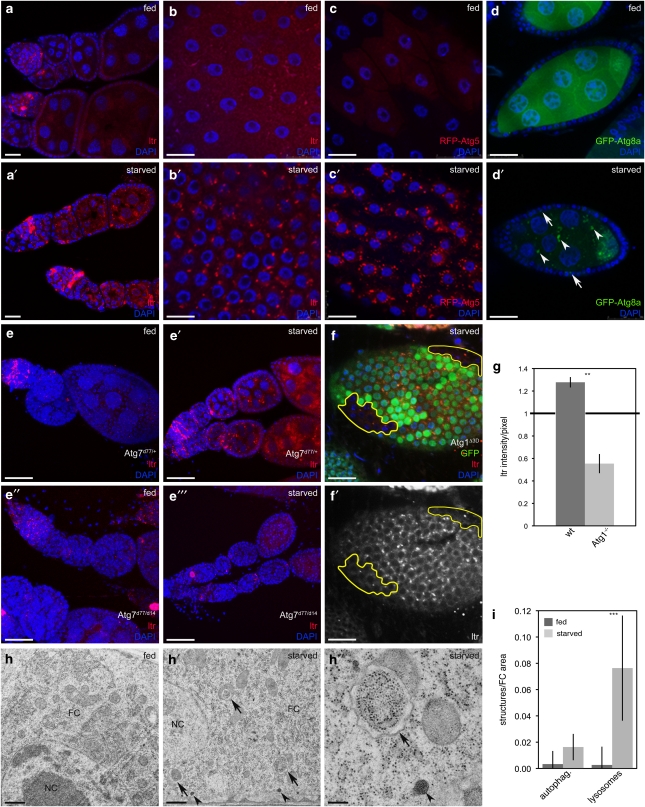
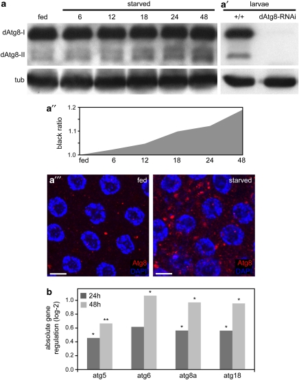
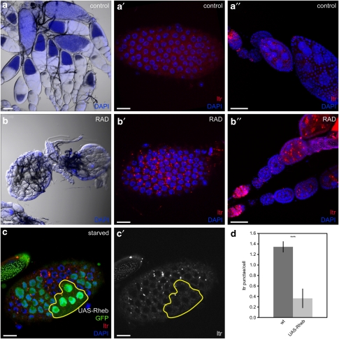
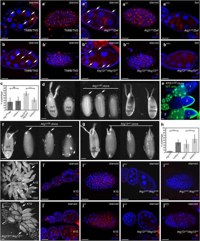
Autophagy, an evolutionarily conserved lysosome-mediated degradation, promotes cell survival under starvation and is controlled by insulin/target of rapamycin (TOR) signaling. In Drosophila, nutrient depletion induces autophagy in the fat body. Interestingly, nutrient availability and insulin/TOR signaling also influence the size and structure of Drosophila ovaries, however, the role of nutrient signaling and autophagy during this process remains to be elucidated. Here, we show that starvation induces autophagy in germline cells (GCs) and in follicle cells (FCs) in Drosophila ovaries. This process is mediated by the ATG machinery and involves the upregulation of Atg genes. We further demonstrate that insulin/TOR signaling controls autophagy in FCs and GCs. The analysis of chimeric females reveals that autophagy in FCs, but not in GCs, is required for egg development. Strikingly, when animals lack Atg gene function in both cell types, ovaries develop normally, suggesting that the incompatibility between autophagy-competent GCs and autophagy-deficient FCs leads to defective egg development. As egg morphogenesis depends on a tightly linked signaling between FCs and GCs, we propose a model in which autophagy is required for the communication between these two cell types. Our data establish an important function for autophagy during oogenesis and contributes to the understanding of the role of autophagy in animal development.


Figures

Figure 1
Figure 1
Starvation induces autophagy in Drosophila FCs and GCs. (a and b) LTR staining is increased in germaria, GCs (a′) and in stage 8 FCs (b′) upon starvation. (c and d) RFP-dAtg5 accumulates upon starvation in stage 8 FCs (c′) and GFP-dAtg8a in FCs (arrows) and GCs (arrowheads) (d′). (ee″′) Atg7 mutants fail to induce autophagy. (f and f′) Atg1 mutant FC clones (marked by the lack of GFP) do not induce LTR staining. (g) LTR intensity/pixel of Atg1 mutant clones normalized to heterozygous cells. (hh″) TEM images depict an accumulation of autophagosomes (arrows) and lysosomes (arrowheads) in starved FCs. (i) TEM quantification of FCs from fed versus starved flies (n=2). Only healthy egg chambers were considered for the analysis. Scale bars: (a, a′, d, d′, f and f′) 20 μm, (bc′) 10 μm, (ee″′) 50 μm, (h and h′) 500 nm, (h″) 200 nm. Error bars show S.D. of the mean, ***P<0.001, **P<0.01.Genotypes: (ab and hh′) y w, (c) da-Gal4/UAS-RFP-dAtg5, (d) da-Gal4/UASp-dAtg8a, (e) Atg7d14/Atg7d77, Atg7d14, (f) hs flp/+Atg1Δ3D FRT80B/FRT80B-UbiGFP
Figure 2
Figure 2
Starvation induces dAtg8 conversion and Atg gene expression in Drosophila ovaries. (a) Western blot (WB) showing the increase of dAtg8-II in a starvation time course. (a′) Expression of dAtg8 is diminished in larvae ubiquitously expressing dAtg8a-RNAi. Tubulin served as loading control. (a″) Quantification of Atg8-II WB signals measured as grey values using ImageJ. Rising grey values represent the increase of dAtg8-II in a. (a″′) Accumulation of dAtg8 labeled autophagosomes in starved stage 8 FCs. (b) Quantitative real-time PCR of ovary RNA samples from fed flies (reference expression level), 24 and 48 h starved flies. n=5; P-values: *P<0.05, **P<0.01. Scale bars: (a″′) 10 μm. Genotypes: (a, a″′ and b) y w, (a′) y w, UAS-dAtg8-RNAi, da-Gal4
Figure 3
Figure 3
IIS/TOR signaling controls autophagy in Drosophila ovaries. (ab″) Injection of RAD leads to small ovaries lacking vitellogenic stages (b) and a strong accumulation of autophagolysosomes in FCs (b′) and GCs (b″). Control ovaries are of normal size (a) and barely show LTR staining in FCs (a′) or GCs (a″). (c and c′) Generation of stage 7 FC clones overexpressing Rheb using the flp-out-Gal4/UAS method results in cells with high (strong GFP signal) and low (weak GFP signal) transgene expression. Only cells with bright GFP signals and enlarged nuclei (as an indication of enhanced cell size due to Rheb overexpression) were considered for the analyses. (d) Quantification of LTR staining in Rheb overexpressing clones compared with WT cells. Error bars show S.D. of the mean, n=8, ***P<0.001. Scale bars: (a and b) 100 μm, (a′, b′, c and c′) 10 μm, (a″ and b″) 50 μm. Genotypes: (ab) y w, (c) hs flp/+act4CD24Gal4 UAS-GFP/UAS-RhebEP50.084
Figure 4
Figure 4
Atg1 and Atg13 are required for FC development. (ac) GC mosaics. Starvation induces autophagy (monitored by LTR staining) in GCs (a and b) and FCs (a′ and b′) of control TM6B/TM3 GC chimeras, whereas GC chimeras homozygous mutant for Atg1 (a″ and a″′) or Atg13 (b″ and b″′) display LTR staining only in WT FCs, which is not seen in fed control GC chimeras (a″″ and b″″). Quantification of offspring shows similar hatching rates as control TM6B/TM3 females (c). (dh) FC mosaics. (d) X-ray induced FC clones. Shown are a normal egg deposited by a WT fly, a flaccid Apc mutant egg, missing DAs and anterior chorion structures, and eggs with Atg1 mutant FC clones, missing DAs, but showing a micropyle (arrows). Generation of WT FC clones in Apc/+ animals completely rescued the Apc phenotype. Typical examples of the resulting WT-like eggs are shown (WT clone). (eh) Heat-shock induced clones. (e) Hs-flp induced mitotic recombination results in ovaries comprising WT (α) or completely mutant (β) egg chambers, egg chambers with all FCs mutant (γ), all GCs mutant (δ) or mosaic FCs (ɛ). (f) Eggs with Atg1 mutant clones lack DAs, but feature a micropyle (arrows). (g) Eggs with Atg13 mutant clones show variable phenotypes with reduced DAs. (h) Hatching rate of eggs containing Atg1 or Atg13 mutant FC clones. (i and j) Ovarian chimeras generated by larval ovary transplantations. After implantation, both K10 and Atg1 (i) or Atg13 (j) mutant ovaries are attached to the oviduct. Upon starvation, autophagy (monitored by LTR staining) is induced in GCs (i′ and j′) and FCs (i″ and j″) of K10 control ovaries, whereas Atg mutant ovaries are unable to induce autophagy (i″′j″″). Error bars show S.D. of the mean, ***P<0.001, **P<0.01. Scale bars: (ab″″, e, i′i″″ and j′j″″) 20 μm, (d, f and g) 100 μm, (i and j) 250 μm. Genotypes: (aa″″) donor: Atg1Δ3D-FRT80B/TM6B, Df(3L)BSC613/TM3, host: Tm2gs/Tm2gs, (bb″″) donor: Atg13Δ81/TM6B, Atg13Δ74/TM3, host: Tm2gs/Tm2gs, (d) w1118, Fs(3)Apc/+, Fs(3)Apc/Atg1Δ3D-FRT80B, (f and e) FRT80isogenic/FRT80-UbiGFP (WT clone), Atg1Δ3D-FRT80B/FRT80-UbiGFP, (g) FRT82isogenic/FRT82-UbiGFP (WT clone), Atg13Δ81-FRT82/FRT82-UbiGFP, (i and j) donor: Atg1Δ3D/Atg1Δ3D, Atg13Δ74/Atg13Δ81 host: fs(1)K10/fs(1)K10

Similar articles

Cited by

References

    1. Melendez A, Neufeld TP. The cell biology of autophagy in metazoans: a developing story. Development. 2008;135:2347–2360. - PMC - PubMed
    1. Neufeld TP. TOR-dependent control of autophagy: biting the hand that feeds. Curr Opin Cell Biol. 2010;22:157–168. - PMC - PubMed
    1. Rusten TE, Lindmo K, Juhasz G, Sass M, Seglen PO, Brech A, et al. Programmed autophagy in the Drosophila fat body is induced by ecdysone through regulation of the PI3K pathway. Dev Cell. 2004;7:179–192. - PubMed
    1. Scott RC, Schuldiner O, Neufeld TP. Role and regulation of starvation-induced autophagy in the Drosophila fat body. Dev Cell. 2004;7:167–178. - PubMed
    1. Drummond-Barbosa D, Spradling AC. Stem cells and their progeny respond to nutritional changes during Drosophila oogenesis. Dev Biol. 2001;231:265–278. - PubMed

Publication types

Substances