Review
. 2011 Jan;10(1):29-46.
doi: 10.1038/nrd3321. Epub 2010 Dec 10.

Ubiquitin-like protein conjugation and the ubiquitin-proteasome system as drug targets

Affiliations
Review

Ubiquitin-like protein conjugation and the ubiquitin-proteasome system as drug targets

Lynn Bedford et al. Nat Rev Drug Discov. 2011 Jan.

Abstract

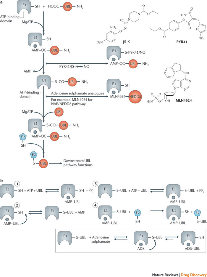
The ubiquitin-proteasome system (UPS) and ubiquitin-like protein (UBL) conjugation pathways are integral to cellular protein homeostasis. The growing recognition of the fundamental importance of these pathways to normal cell function and in disease has prompted an in-depth search for small-molecule inhibitors that selectively block the function of these pathways. However, our limited understanding of the molecular mechanisms and biological consequences of UBL conjugation is a significant hurdle to identifying drug-like inhibitors of enzyme targets within these pathways. Here, we highlight recent advances in understanding the role of some of these enzymes and how these new insights may be the key to developing novel therapeutics for diseases including immuno-inflammatory disorders, cancer, infectious diseases, cardiovascular disease and neurodegenerative disorders.


Conflict of interest statement

Lawrence R. Dick and James E. Brownell are employees of Millennium Pharmaceuticals. Lynn Bedford, James Lowe and R. John Mayer declare no competing financial interests.

Figures

Figure 1
Figure 1. Overview of the enzymatic cascade involved in ubiquitin-like protein (UBL) conjugation and the ubiqitin–proteasome system (UPS).
a | Ubiquitin-activating enzyme (UAE) binds ATP and ubiquitin (Ub) to form a ternary complex consisting of E1–ubiquitin thioester with ubiquitin–AMP bound (see text for details). The thioester-bound ubiquitin is then passed to one of several E2 conjugating enzymes through a transthiolation reaction. The ubiquitin-charged E2 then forms a complex with an E3 ligase and a protein substrate to transfer ubiquitin to a lysine residue on the substrate. To mark substrates for degradation, multiple ubiquitins are similarly recruited to produce a K48-linked polyubiquitin chain. Following release from the E3, the proteasome recognizes the polyubiquitin chain, and the substrate is deubiquitylated and destroyed. Substrates marked with ubiquitin chains linked through lysines 6, 11, 27, 29 and 33 also seem to be primarily destined for degradation. Alternatively, substrates marked with monoubiquitin, linear ubiquitin chains or K63 ubiquitin chains are involved in signalling functions that are independent of the proteasome. A second E1 (ubiquitin-like modifier activating enzyme 6 (UBA6)) also activates ubiquitin, but the function(s) of this pathway are unknown. b | Nine classes of UBL and eight E1 activating enzymes participate in diverse biological pathways in humans. E1s, E2s and UBLs are structurally and mechanistically related but are unique to each pathway. Ubiquitin and some E2s are exceptions in that they can be used by both the UAE and UBA6 pathways. Ufl1 and C20orf166 were recently identified and reported by Tatsumi et al. DUB, deubiquitylating enzyme; NAE, NEDD8-activating enzyme; PPi, inorganic pyrophosphate; SAE, SUMO-activating enzyme.
Figure 2
Figure 2. Mechanisms of E1 inhibitors identified in studies of different ubiquitin-like protein (UBL) pathways.
a | In the first step of UBL activation, E1s bind ATP and a cognate UBL and catalyse the formation of a UBL carboxy- terminal acyl adenylate. The E1 catalytic cysteine then attacks the UBL–adenylate to form a thioester with the C terminus of the UBL. The E1 subsequently binds a second ATP and UBL, again forming a UBL–adenylate and resulting in the formation of a ternary complex consisting of an E1–UBL thioester with UBL–adenylate bound to it. This form of the E1 is fully competent to transfer thioester-bound UBL to a cognate E2 enzyme and initiate the downstream effects of UBL signalling. Small molecules, including the pyrazone derivative PYR41, JS-K and MLN4924, use distinct mechanisms to block this process at different stages of the E1 reaction cycle (see Box 2 for further details). b | Substrate-assisted mechanism-based E1 inhibition. E1s use a multistep mechanism to form a ternary complex consisting of an E1∼UBL thioester (∼ denotes a high-energy bond) with UBL–adenylate bound to it (steps 1–3). This form of E1 is competent for UBL transfer to an E2 by a transthiolation reaction (step 4). The NEDD8-activating enzyme (NAE)-selective inhibitor MLN4924 and related adenosine sulphamate analogues are mechanism-based inhibitors of E1s and form covalent UBL–inhibitor adducts in situ, catalysed by the E1 itself. Inhibitors of this class bind exclusively to the UBL thioester form of E1 shown in step 2 and attack the thioester bond to yield the UBL–inhibitor adduct. The UBL–inhibitor adduct mimics UBL–adenylate, the first intermediate in the E1 reaction cycle, but cannot be further used in subsequent intra-enzyme reactions. The stability of the UBL–inhibitor adduct within the E1 active site adenylation domain blocks enzyme activity.
Figure 4
Figure 4. Molecular interactions of E2s suggest potential modes of inhibition.
a | E2s engage in a sequence of highly specific interactions to faithfully transfer a ubiquitin-like protein (UBL) to a substrate. E2s first bind their respective E1 to receive an activated UBL through a transthiolation reaction. In most cases, the E2–UBL thioester then binds a specific E3 or E3–substrate complex. During catalysis to protein substrates, the UBL is transferred from the E2 thiol active site to the amino group of a substrate acceptor lysine residue that is positioned for nucleophilic attack on the E2–UBL thioester bond. Each of these E2 interactions offers potential opportunities for selective inhibition as illustrated by the following examples. b | Targeting E2∼UBL thioester formation by blocking E1–E2 protein–protein interaction. A synthetic peptide called UBC12N26 corresponds to the 26-residue amino terminus extension of UBC12 that specifically binds the NEDD8-activiating enzyme (NAE). UBC12N26 competes for binding of UBC12 to NAE, thereby blocking transthiolation of NEDD8 to UBC12 and inhibiting downstream NEDD8-dependent events. c | Targeting E3-dependent E2 allosteric activation. In some cases, E2–E3 binding is necessary but not sufficient for optimal UBL transfer to a substrate. For example, the E3 gp78 binds the E2, UBE2G2, through a RING finger and a second domain called G2BR that binds the backside of UBE2G2, which is opposite from the catalytic cysteine and distal from the RING binding surface. Blocking G2BR interaction with UBE2G2 would inhibit allosteric activation of the E2 and prevent subsequent ubiquitylation of substrate proteins. d | Targeting catalysis of UBL transfer. A conserved asparagine residue in E2s (exemplified by asparagine 79 in UBC13) has been shown to be crucial for UBL transfer and is thought to form the oxyanion hole required to stabilize the E2 thioester–substrate transition state intermediate. Targeting this site with a small-molecule effector may offer yet another approach for inhibition. Ub, ubiquitin. Part b is reproduced, with permission, from Ref. © (2004) Macmillan Publishers Ltd. All rights reserved. Part c is reproduced, with permission, from Ref. © (2009) Elsevier Science. Part d is reproduced, with permission, from Ref. © (2003) Macmillan Publishers Ltd. All rights reserved.
Figure 3
Figure 3. Structural differences among various types of E3 ligases.
There are four major classes of ubiquitin ligase: HECT domain proteins, U-box proteins, monomeric RING finger E3s, and multisubunit E3 complexes that contain a RING finger protein. a | HECT domain E3s use a unique mechanism in which ubiquitin (Ub) is transferred from an E2 to a conserved cysteine residue within the E3 via transthiolation and is then transferred from the E3 to a substrate amino group. All other types of E3 facilitate transfer of Ub from a charged E2 directly to a substrate. b | The RING finger motif comprises a Zn2+ binding domain that is required for E2 binding. The U-box also binds E2s and is structurally similar to the RING motif, but does not bind metal ions. Monomeric RING finger E3s and U-box E3s bind to both the substrate and the E2. In multimeric RING finger complexes, the RING finger protein binds the E2 while other proteins in the complex bind the substrate. These multimeric complexes include the anaphase promoting complex/cyclosome and cullin-RING ligases (CRLs). A unique aspect of CRLs is the requirement for modification of the cullin subunit by NEDD8 for ubiquitin ligase activity (Box 2). RBX, RING-box protein.
Figure 5
Figure 5. Ubiquitin and signalling to nuclear factor-κB (NF-κB).
a | Tumour necrosis factor-α (TNF-α) signalling. b | Interleukin-1 (IL-1) receptor 1 (IL1R1) and Toll-like receptor signalling. The diagrams are based on recent reviews,. Although it is tempting to represent a general consensus, some enzymological steps are controversial due, for example, to enzymological redundancy. The role of ubiquitin (Ub) in signalling to NF-κB needs considerably more investigation. See the main text for more details. DUB, deubiquitylating enzyme; LUBAC, linear ubiquitin chain-assembly complex.
Figure 6
Figure 6. Protein quality control and neurodegeneration.
Neuronal proteins are normally degraded by the activities of the ubiquitin–proteasome system (UPS), chaperone-mediated autophagy (CMA) and macro-autophagy (collectively referred to as autophagy), and the endosome–lysosome pathway. Unfolded proteins, proteins altered by mutation or post-translational modifications and proteins that are damaged (for example, by oxidative stress, irradiation or toxins) are recognized by molecular chaperones and delivered to the UPS and autophagy pathways. Age-related and chronic neurodegenerative diseases, including Alzheimer's disease, Parkinson's disease, dementia with Lewy bodies and amyotrophic lateral sclerosis, are 'proteinopathies' associated with the intraneuronal accumulation of insoluble protein aggregates resulting from the malfunction of the neuronal UPS and/or autophagy pathways (see main text for details). The mechanisms of how neurodegeneration results from abnormal protein accumulation due to impaired function of the UPS and autophagy pathways are not known.

References

    1. Ciechanover A, Schwartz AL. The ubiquitin-proteasome pathway: the complexity and myriad functions of proteins death. Proc. Natl Acad. Sci. USA. 1998;95:2727–2730. - PMC - PubMed
    1. Hoeller D, Dikic I. Targeting the ubiquitin system in cancer therapy. Nature. 2009;458:438–444. - PubMed
    1. Lehman NL. The ubiquitin proteasome system in neuropathology. Acta Neuropathol. 2009;118:329–347. - PMC - PubMed
    1. Corn PG. Role of the ubiquitin proteasome system in renal cell carcinoma. BMC Biochem. 2007;8:S4. - PMC - PubMed
    1. Herrmann J, Ciechanover A, Lerman LO, Lerman A. The ubiquitin–proteasome system in cardiovascular diseases-a hypothesis extended. Cardiovasc. Res. 2004;61:11–21. - PubMed

Publication types