. 2011 Jan;13(1):13-21.
doi: 10.1038/ncb2135. Epub 2010 Dec 12.

LIF-independent JAK signalling to chromatin in embryonic stem cells uncovered from an adult stem cell disease

Affiliations

LIF-independent JAK signalling to chromatin in embryonic stem cells uncovered from an adult stem cell disease

Dean S Griffiths et al. Nat Cell Biol. 2011 Jan.

Abstract

Activating mutations in the tyrosine kinase Janus kinase 2 (JAK2) cause myeloproliferative neoplasms, clonal blood stem cell disorders with a propensity for leukaemic transformation. Leukaemia inhibitory factor (LIF) signalling through the JAK-signal transducer and activator of transcription (STAT) pathway enables self-renewal of embryonic stem (ES) cells. Here we show that mouse ES cells carrying the human JAK2V617F mutation were able to self-renew in chemically defined conditions without cytokines or small-molecule inhibitors, independently of JAK signalling through the STAT3 or phosphatidylinositol-3-OH kinase pathways. Phosphorylation of histone H3 tyrosine 41 (H3Y41) by JAK2 was recently shown to interfere with binding of heterochromatin protein 1α (HP1α). Levels of chromatin-bound HP1α were lower in JAK2V617F ES cells but increased following inhibition of JAK2, coincident with a global reduction in histone H3Y41 phosphorylation. JAK2 inhibition reduced levels of the pluripotency regulator Nanog, with a reduction in H3Y41 phosphorylation and concomitant increase in HP1α levels at the Nanog promoter. Furthermore, Nanog was required for factor independence of JAK2V617F ES cells. Taken together, these results uncover a previously unrecognized role for direct signalling to chromatin by JAK2 as an important mediator of ES cell self-renewal.


Figures
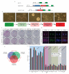
Figure 1
Figure 1
JAK2V617F sustains ES cells in a self-renewing state without any additional factors a. Targeting strategy to insert patient cDNA containing JAK2V617F mutation into the jak2 allele by homologous recombination. b. JAK2V617F ES cells are made factor-independent by transferring ES cells growing in N2B27 plus LIF and BMP4 into N2B27 only. The ES cells then undergo a crisis; detaching and forming spheres, these spheres can then be reattached by transferring into fresh N2B27 on freshly gelatinised flasks. c. ES cells were plated at 1 × 10³ cells per well of a 12 well plate, 6 days later the cells were fixed and stained for alkaline phosphatase to identify ES cell colonies. Only factor-independent JAK2V617F ES cells (JAK2V617F) can form colonies in N2B27 only, this ability is lost following the addition of AG490. d. Parental ES cells grown in N2B27 plus LIF and BMP4 have a similar pattern of staining for the key ES cell transcription factors Nanog and Oct4 to factor-independent JAK2V617F ES cells in N2B27 alone. Of note, factor-independent JAK2V617F cells have the characteristic variable levels of Nanog seen with wild type ES cells. Scale bar 20μm. e. Microarray analysis of the three ES cell lines demonstrates that the majority of expressed genes are common to all three ES cell lines. Correlation coefficients were calculated using the mean of each two-way comparisons and show a strong correlation coefficient between all three datasets (red values). f. Genes known to be critical for ES cell self-renewal are expressed at similar levels in all datasets, and there is no up-regulation of genes expressed in more differentiated cell types. Values are mean of three biological replicates, error bars represent S.E.M.
Figure 2
Figure 2
Factor independent JAK2V617 ES cells are capable of multilineage differentiation in vitro and in vivo a. Factor independent JAK2V617F ES cells give rise to embryoid bodies containing red blood cells and differentiate into neurons when transferred into appropriate differentiation conditions. b. JAK2V617F ES cells generate Flk-1 positive mesodermal cells with delayed differentiation kinetics. Wild type and factor-independent JAK2V617F ES cells were differentiated to haematopoietic lineages, and the proportion of Flk-1 and SSEA-1 positive cells were measured by FACS at days 3, 5 and 7 of differentiation c. Teratocarcinoma formation from JAK2V617F ES cells. Parental AB2.2 ES cells maintained in N2B27 plus LIF and BMP4 and factor-independent JAK2V617F ES cells were injected into the kidney capsule of 129sv mice and left for four weeks. Teratocarcinomas consisting of cells from all three germ layers formed, but the majority the teratocarcinomas from factor-independent JAK2V617F ES cells remained undifferentiated or were of no immediately discernible cell type. Un -undifferentiated, ec- ectoderm, me – mesoderm, en – endoderm.
Figure 3
Figure 3
JAK2V617F does not activate canonical signalling pathways independently of cytokines, but JAK is required for ES cell self-renewal. a. Colony forming assay for wild type and factor-independent ES cells in 2i or N2B27 alone following clonal growth for 6 days in dilution series of JAK inhibitors. ES colonies confirmed by positive staining for alkaline phosphatase. There was a significant reduction (p<1×10⁻⁷) for all inhibitors on all cell types except JAK2V617F ES cells in N2B27 alone at the lowest concentrations of TG101209, for details of GLM see Supplementary table 1. Error bars represent S.E.M. b. Immunoblot analysis of steady state levels, or following 8 hours of treatment of cells with LIF or AG490 for P-STAT3 (tyr705), STAT3, P-AKT (ser 473) and tubulin of wild type ES cells grown in N2B27 plus 2i, JAK2V617F ES cells in N2B27 plus 2i or JAK2V71F ES cells in N2B27 only. c. Colony forming assay for STAT3 null ES cells in 2i performed as in 3c. There was a significant reduction (p<1 × 10⁻¹⁶) using all inhibitors. For details of GLM see Supplementary table 1. d. Immunocytochemistry of STAT3 null JAK2V617F factor-independent ES cells confirms continued expression of key ES cell genes Oct4 and Nanog. Scale bar 20μm.
Figure 4
Figure 4
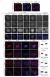
JAK2 is present in the nucleus of ES cells and JAK2 dynamically regulates HP1α access to chromatin by phosphorylating H3Y41. a. Immunohistochemistry for phosphorylated JAK2 in wild type ES cells growing in 2i. Orthogonal view confirms the presence of phosphorylated JAK2 in the nucleus. Scale bar 20μm. b. Immunohistochemistry confirms that HP1α was present at lower levels in JAK2V617F ES cells compared to parental cells when they are maintained in multiple ES cell conditions, but Oct4 remains unchanged. Scale bar 20μm. c. Immunohistochemistry for HP1α and Nanog in steady state factor-independent JAK2V617F ES cells and following treatment with TG101209 for 2 hours. There was a significant increase in the level of HP1α and decrease in Nanog following inhibitor treatment, two independent experiments combined in box and whisker plot, difference determined by Students T-Test. N is cell number. Scale bar 20μm. d. Immunohistochemistry for H3Y41ph and Nanog in steady state factor-independent JAK2V617F ES cells and following treatment with TG101209 for 2 hours. There was a significant decrease in the levels of H3Y41ph and Nanog following inhibitor treatment, two independent experiments combined in box and whisker plot, difference determined by Students T-Test. N is cell number. Scale bar 20μm.
Figure 5
Figure 5
JAK2 regulates H3Y41ph at the Nanog promoter and Nanog is critical for factor-independent self-renewal. a. A 8kb window surrounding the Nanog TSS was interrogated using chromatin immunoprecipitation for H3Y41ph in steady state factor-independent JAK2V617F ES cells or following treatment with TG101209 for 6 hours. Data were normalised to H3 occupancy. Representative plot of two independent experiments, error bars represent S.E.M. b. The Nanog promoter was interrogated using chromatin immunoprecipitation for H3K4me3, H3Y41ph and HP1α in wild type ES cells growing in N2B27 plus 2i or following treatment with AG490 for 16 hours. Data were normalised to H3 occupancy. Representative plot of two independent experiments, error bars represent S.E.M c. Colony forming assay for Nanog over-expressing ES cells, performed as described in 3b. While JAK inhibition caused a decrease in ES cell self-renewal of Nanog over-expressing ES cells, this was significantly less than wild type ES cells, * p<0.05, ** p<0.01, ***p<0.001, see Supplementary table 1 for details of T-Test. Error bars represent S.E.M. d. Growth of JAK2V617F ES cells in N2B27 alone is compromised following transduction with a shRNA Nanog knockdown lentivirus co-expressing eGFP. e. Immunohistochemistry of JAK2 Null ES cells growing in 2i media demonstrated that H3Y41 is phosphorylated in the absence of JAK2, which in turn was dynamically regulated by JAK inhibition treatment with AG490 for 16 hours. There was a significant decrease in the levels of H3Y41ph and Oct4 following inhibitor treatment, two independent experiments combined in box and whisker plot, difference determined by Students T-Test. N is cell number.
Figure 6
Figure 6
JAK2 is not the only JAK which can phosphorylate H3Y41 a. Immunohistochemistry of JAK2 Null ES cells growing in 2i media demonstrated that H3Y41 is phosphorylated in the absence of JAK2, which in turn was dynamically regulated by JAK inhibition treatment with AG490 for 16 hours. There was a significant decrease in the levels of H3Y41ph and Oct4 following inhibitor treatment, two independent experiments combined in box and whisker plot, difference determined by Students T-Test. N is cell number. b. Orthogonal view of immunohistochemistry for phosphorylated JAK1 in wild type ES cells maintained in N2B27 plus 2i.

References

    1. Campbell PJ, Green AR. The myeloproliferative disorders. N. Engl. J. Med. 2006;355:2452–2466. - PubMed
    1. Levine RL, Gilliland DG. Myeloproliferative disorders. Blood. 2008;112:2190–2198. - PMC - PubMed
    1. James C, et al. A unique clonal JAK2 mutation leading to constitutive signalling causes polycythaemia vera. Nature. 2005;434:1144–1148. - PubMed
    1. Levine RL, et al. Activating mutation in the tyrosine kinase JAK2 in polycythemia vera, essential thrombocythemia, and myeloid metaplasia with myelofibrosis. Cancer Cell. 2005;7:387–397. - PubMed
    1. Baxter EJ, et al. Acquired mutation of the tyrosine kinase JAK2 in human myeloproliferative disorders. Lancet. 2005;365:1054–1061. - PubMed

Publication types

MeSH terms