. 2010 Sep 24:4:176.
doi: 10.3389/fnhum.2010.00176. eCollection 2010.

Face-Specific Resting Functional Connectivity between the Fusiform Gyrus and Posterior Superior Temporal Sulcus

Affiliations

Face-Specific Resting Functional Connectivity between the Fusiform Gyrus and Posterior Superior Temporal Sulcus

Nicholas B Turk-Browne et al. Front Hum Neurosci. .

Abstract

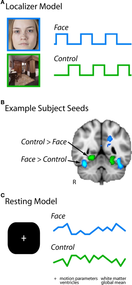
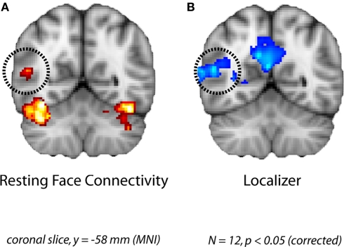
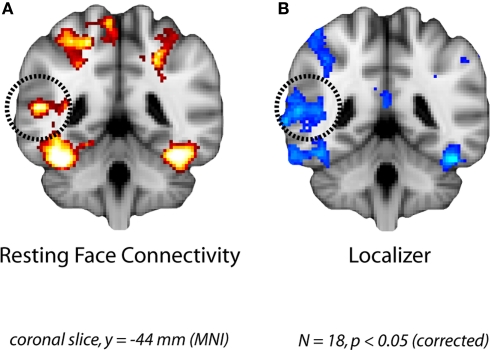
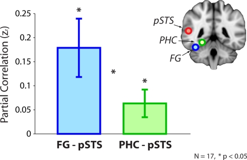
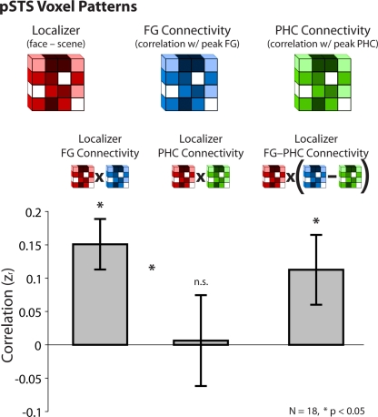
Faces activate specific brain regions in fMRI, including the fusiform gyrus (FG) and the posterior superior temporal sulcus (pSTS). The fact that the FG and pSTS are frequently co-activated suggests that they may interact synergistically in a distributed face processing network. Alternatively, the functions implemented by these regions may be encapsulated from each other. It has proven difficult to evaluate these two accounts during visual processing of face stimuli. However, if the FG and pSTS interact during face processing, the substrate for such interactions may be apparent in a correlation of the BOLD timeseries from these two regions during periods of rest when no faces are present. To examine face-specific resting correlations, we developed a new partial functional connectivity approach in which we removed variance from the FG that was shared with other category-selective and control regions. The remaining face-specific FG resting variance was then used to predict resting signals throughout the brain. In two experiments, we observed face-specific resting functional connectivity between FG and pSTS, and importantly, these correlations overlapped precisely with the face-specific pSTS region obtained from independent localizer runs. Additional region-of-interest and pattern analyses confirmed that the FG-pSTS resting correlations were face-specific. These findings support a model in which face processing is distributed among a finite number of connected, but nevertheless face-specialized regions. The discovery of category-specific interactions in the absence of visual input suggests that resting networks may provide a latent foundation for task processing.

Keywords: fMRI; face processing; functional connectivity; fusiform face area; high-level vision; inferior temporal cortex; rest; scene processing.


Figures

Figure 1
Figure 1
Analysis pipeline. (A) Localizer runs were modeled with regressors for face blocks and control blocks (scenes or flowers in Experiment 1, scenes in Experiment 2). (B) The contrast of these two regressors identified two seed regions in each subject for further investigation: the right fusiform gyrus (FG), and an adjacent region of right ventral temporal cortex selective for flowers or scenes (the latter is the PHC). (C) Timeseries were extracted from these regions in each resting run, and entered simultaneously in a GLM of the same run (along with other nuisance regressors of no interest).
Figure 2
Figure 2
Face-specific connectivity from Experiment 1. (A) Group analysis of resting correlations with each subject's face-specific FG after removing variance from the control region. (B) The pSTS region that was correlated with the FG at rest overlapped with the face-selective pSTS region obtained in a group analysis of the localizer runs.
Figure 3
Figure 3
Face-specific connectivity from Experiment 2. (A) Group analysis of resting correlations with each subject's face-specific FG after removing variance from their scene-specific PHC. (B) The pSTS region that was correlated with the FG at rest overlapped with the face-selective pSTS region obtained in a group analysis of the localizer runs.
Figure 4
Figure 4
ROI analysis from Experiment 2. Resting correlations between the FG (after partialing out the PHC) and the pSTS, and the PHC (after partialing out the FG) and the pSTS. Correlations were computed within-subject using peak voxels from the localizer and converted to z-scores for statistical tests. Error bars reflect standard errors of the mean.
Figure 5
Figure 5
Pattern analysis from Experiment 2. Across a population of pSTS voxels, correlations between the strength of face-selectivity from the localizer and the strength of resting connectivity with the peak FG (after partialing out the PHC) and the peak PHC (after partialing out the FG). Voxels that were more face-selective in the localizer were also more correlated with the FG at rest, but there was no such relationship in either direction for the PHC. Moreover, face-selectivity in the localizer (face–scene) predicted face-selectivity in partial resting connectivity (FG–PHC connectivity). Correlations were computed within-subject and converted to z-scores for statistical tests. Error bars reflect standard errors of the mean.
Figure A1
Figure A1
Example resting run model from Experiment 1. Resting timeseries were extracted from the localized FG and control seed regions, and entered into a multiple regression model of the same resting run with several nuisance regressors of no interest.
Figure A2
Figure A2
Group confusion matrix from Experiment 1. Crosscorrelation of all regressors from resting run models for each subject (concatenating regressors across runs within subject), converted to z-scores using Fisher’s r-to-z transformation, and evaluated across subjects with one-sample t-tests.
Figure A3
Figure A3
Effect of kernel in Experiment 2. (A) Group analysis of resting correlations with each subject’s peak face-selective FG voxel after removing variance from the peak scene-selective PHC voxel. (B) Group analysis of resting correlations when an 8-mm FWHM Gaussian kernel centered on the peak voxel was used to define the seed and control timeseries.

References

    1. Aguirre G. K., Zarahn E., D'Esposito M. (1998). An area within human ventral cortex sensitive to “building” stimuli: evidence and implications. Neuron 21, 373–38310.1016/S0896-6273(00)80546-2 - DOI - PubMed
    1. Allison T., McCarthy G., Nobre A., Puce A., Belger A. (1994). Human extrastriate visual cortex and the perception of faces, words, numbers, and colors. Cereb. Cortex 4, 544–55410.1093/cercor/4.5.544 - DOI - PubMed
    1. Allison T., Puce A., McCarthy G. (2000). Social perception from visual cues: Role of the pSTS region. Trends Cogn. Sci. 4, 267–27810.1016/S1364-6613(00)01501-1 - DOI - PubMed
    1. Allison T., Puce A., Spencer D. D., McCarthy G. (1999). Electrophysiological studies of human face perception. I: Potentials generated in occipitotemporal cortex by face and non-face stimuli. Cereb. Cortex 9, 415–43010.1093/cercor/9.5.415 - DOI - PubMed
    1. Andrews T. J., Ewbank M. P. (2004). Distinct representations for facial identity and changeable aspects of faces in the human temporal lobe. NeuroImage 23, 905–91310.1016/j.neuroimage.2004.07.060 - DOI - PubMed

LinkOut - more resources