

Adenomatoid tumor of testis
- PMID: 21151545
- PMCID: PMC2990235
- DOI: 10.4137/cpath.s3091
Adenomatoid tumor of testis
Abstract
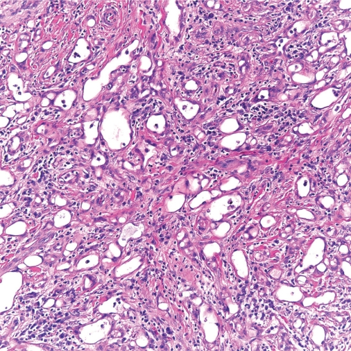
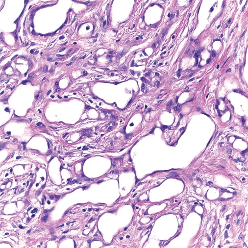
Adenomatoid tumors are responsible for 30% of all paratesticular masses. These are usually asymptomatic, slow growing masses. They are benign tumors comprising of cords and tubules of cuboidal to columnar cells with vacuolated cytoplasm and fibrous stroma. They are considered to be of mesothelial origin supported by histochemical studies and genetic analysis of Wilms tumor 1 gene expression. Excision biopsy is both diagnostic and therapeutic procedure. The main clinical consideration is accurate diagnosis preventing unnecessary orchiectomy. Diagnostic studies include serum tumor markers (negative alpha fetoprotein, beta HCG, LDH) ultrasonography (hypoechoic and homogenous appearance) and frozen section.
Keywords: adenomatoid tumor; paratesticluar masses.
Figures

References
-
- Delahunt B, Eble JN, King D, Bethwaite PB, Nacey JN, Thornton A. Immunohistochemical evidence for mesothelial origin of paratesticular adenomatoid tumour. Histopathology. 2000;36(2):109–15. - PubMed
-
- Eble JN SG, Epstein JI, Sesterhenn IA.World Health Organization Classification of TumorsPthology and Genetics of Tumour of the Urinary System: IACR Press; Lyon: 2004
-
- Stephenson TJ, Mills PM. Adenomatoid tumours: an immunohistochemical and ultrastructural appraisal of their histogenesis. J Pathol. 1986;148(4):327–35. - PubMed
-
- Schwartz EJ, Longacre TA. Adenomatoid tumors of the female and male genital tracts express WT1. Int J Gynecol Pathol. 2004;23(2):123–8. - PubMed
-
- Gokce G, Kilicarslan H, Ayan S, Yildiz E, Kaya K, Gultekin EY. Adenomatoid tumors of testis and epididymis: a report of two cases. Int Urol Nephrol. 2001;32(4):677–80. - PubMed
LinkOut - more resources
Full Text Sources
