. 2010 Dec 7:16:2605-11.

Circadian regulation of the PERIOD 2::LUCIFERASE bioluminescence rhythm in the mouse retinal pigment epithelium-choroid

Affiliations

Circadian regulation of the PERIOD 2::LUCIFERASE bioluminescence rhythm in the mouse retinal pigment epithelium-choroid

Kenkichi Baba et al. Mol Vis. .

Abstract

Purpose: The retinal pigment epithelium (RPE) plays an important role in the maintenance of the health and function of photoreceptors. Previous studies have shown that the RPE is also involved in the regulation of disc shedding, a process that is vital for photoreceptor health. This process has been shown to be under circadian control, although the mechanisms that control it are poorly understood. The aim of the present study was to investigate Period 2 (Per2) mRNA levels in the mouse RPE in vivo, and to determine whether the cultured RPE-choroid from PERIOD 2::LUCIFERASE (PER2::LUC) knockin mice expresses a circadian rhythm in bioluminescence.

Methods: Per2 mRNA levels were measured using real-time quantitative RT-PCR, and bioluminescence was measured in PER2::LUC knockin mice using a Lumicycle®.

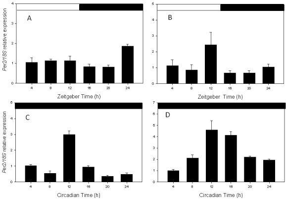
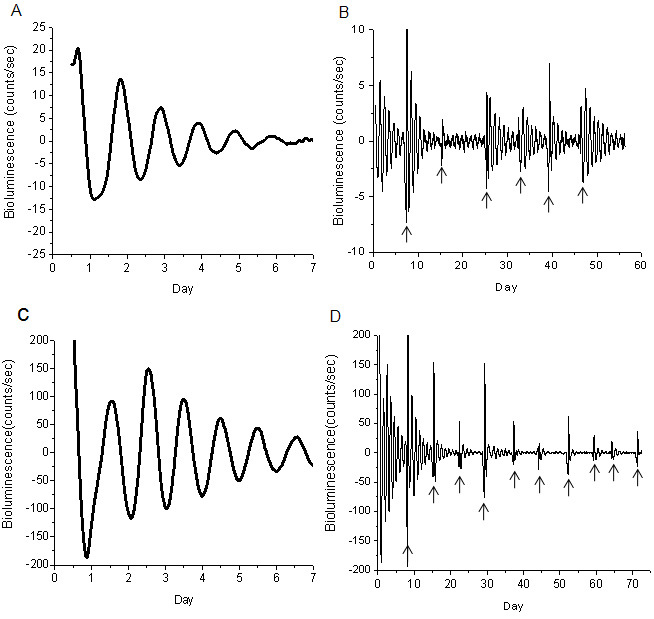
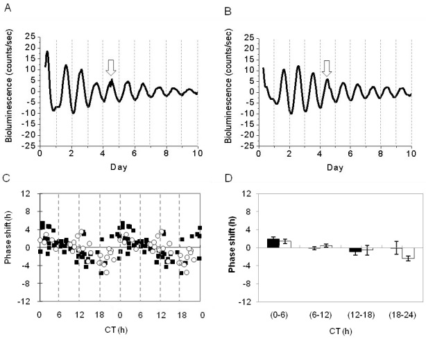
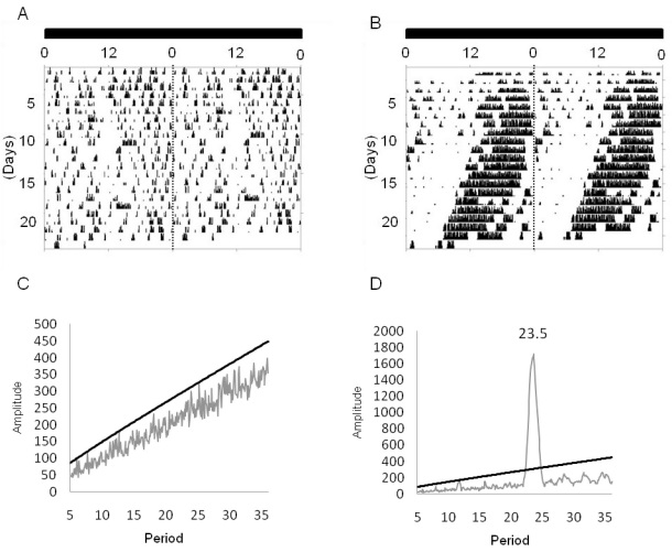
Results: Per2 mRNA levels in the RPE-choroid show a clear circadian rhythm in vivo. A circadian rhythm in PER2::LUC bioluminescence was recorded from cultured RPE-choroid explants. Light exposure during the subjective night did not cause a circadian rhythm phase-shift of PER2::LUC bioluminescence. Finally, removal of the suprachiasmatic nuclei of the hypothalamus did not affect the bioluminescence circadian rhythm in the RPE-choroid.

Conclusions: Our results demonstrate that the RPE-choroid contains a circadian clock, and the regulation of this circadian rhythm resides within the eye. These new data indicate that it may be useful to design studies with the aim of elucidating the molecular mechanisms responsible for the regulation of the rhythmic event in the RPE.


Figures

Figure 1
Figure 1
Per2 circadian expression in retina and retinal pigment epithelium (RPE). A: RPE in Light/Dark cycle. B: Retina in Light/Dark cycle. C: RPE in constant darkness. D: Retina in constant darkness. In both tissues, the Per2 mRNA levels showed a significant circadian rhythm (one-way ANOVA, p<0.05 in all cases). Each point represents the mean±SEM (n=3–7).
Figure 2
Figure 2
PER 2::LUC bioluminescence rhythms in RPE-choroid and retina. A: Representative example of PER2::LUC rhythms in RPE-choroid. B: The circadian rhythms of RPE-choroid persisted more than 50 days. C: Representative example of PER2::LUC rhythm in retina. D: The circadian rhythms of retina persisted more than 70 days. Although the circadian rhythm in the bioluminescence damped out after 6–7 days, a medium exchange could reinitiate the circadian rhythm in these cultures. The arrows indicate the days on which the medium in the culture was replaced.
Figure 3
Figure 3
Light responsiveness of PER2::LUC bioluminescence rhythm in retinal pigment epithelium (RPE)-choroid. A: The light pulse was given at Day 4 of PER2::LUC rhythms. B: The control pulse was given at Day 4 of PER2::LUC rhythms. Arrows indicate the times of the treatment. C: The double-plotted phase response curve to light stimuli in the PER2::LUC bioluminescence rhythm in the RPE-choroid. Black squares indicate control and white circles indicate the samples that received the light pulse. D: Data were divided into 4 bins in 6 h intervals for statistical analysis. No significant differences were observed between the control (black bars) and the experimental (white bars) groups (two-way ANOVA, p>0.1).
Figure 4
Figure 4
Circadian rhythm of locomotor activity in SCN lesioned mice. A: Representative actogram of a mouse that received SCN lesion. B: A mouse that received a sham lesion. C: The periodogram analysis (χ2) indicated that the locomotor activity is no longer rhythmic after the SCN has been lesioned. D: It is still rhythmic in the sham-lesioned mice.
Figure 5
Figure 5
Effect of SCN lesion on the RPE-choroid circadian rhythm. A: Representative example of the RPE-choroid PER2::LUC bioluminescence rhythm in a sham-lesioned mouse B: In a SCN-lesioned mouse. C: Removal of the SCN did not affect the phase of the PER2::LUC RPE-choroid circadian rhythm (white square represents sham-lesioned mice; black square represents SCN-lesioned mice) D: Period of the circadian expression (white bars represents sham operated mice; black bars represents SCN-lesioned mice; t-tests, p>0.1 in both cases).

References

    1. Tosini G, Pozdeyev N, Sakamoto K, Iuvone PM. The circadian clock system in the mammalian retina. Bioessays. 2008;30:624–33. - PMC - PubMed
    1. Tosini G, Menaker M. The clock in the mouse retina: melatonin synthesis and photoreceptor degeneration. Brain Res. 1998;789:221–8. - PubMed
    1. Sakamoto K, Liu C, Kasamatsu M, Iuvone PM, Tosini G. Intraocular injection of kainic acid does not abolish the circadian rhythm of arylalkylamine N-acetyltransferase mRNA in rat photoreceptors. Mol Vis. 2006;12:117–24. - PubMed
    1. Tosini G, Davidson AJ, Fukuhara C, Kasamatsu M, Castanon-Cervantes O. Localization of a circadian clock in mammalian photoreceptors. FASEB J. 2007;21:3866–71. - PMC - PubMed
    1. Cahill GM, Besharse JC. Circadian clock functions localized in xenopus retinal photoreceptors. Neuron. 1993;10:573–7. - PubMed

Publication types

MeSH terms

LinkOut - more resources