. 2010 Dec 3:16:2539-49.

Spatiotemporal expression pattern of ceramide kinase-like in the mouse retina

Affiliations

Spatiotemporal expression pattern of ceramide kinase-like in the mouse retina

Sharon Vekslin et al. Mol Vis. .

Abstract

Purpose: The CERKL gene encodes for ceramide kinase-like, a novel protein of unknown function. CERKL mutations are associated with a severe retinal phenotype. The purpose of this work was to investigate alternative splicing, and the temporal and spatial expression pattern of CERKL in the mouse retina.

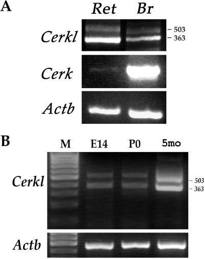
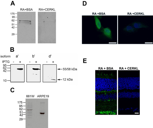
Methods: Reverse Transcription-Polymerase Chain Reaction (RT-PCR) analysis of mouse retina RNA was used to study the expression of Cerkl at various developmental time points, and to identify its various splice-isoforms. A specific anti-CERKL antibody was developed and used for immunostaining to study the localization of the endogenous CERKL protein in retina-derived cell lines and in the mouse retina.

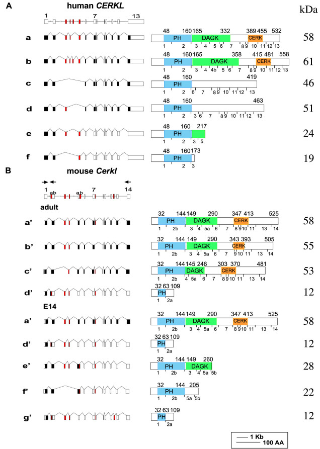
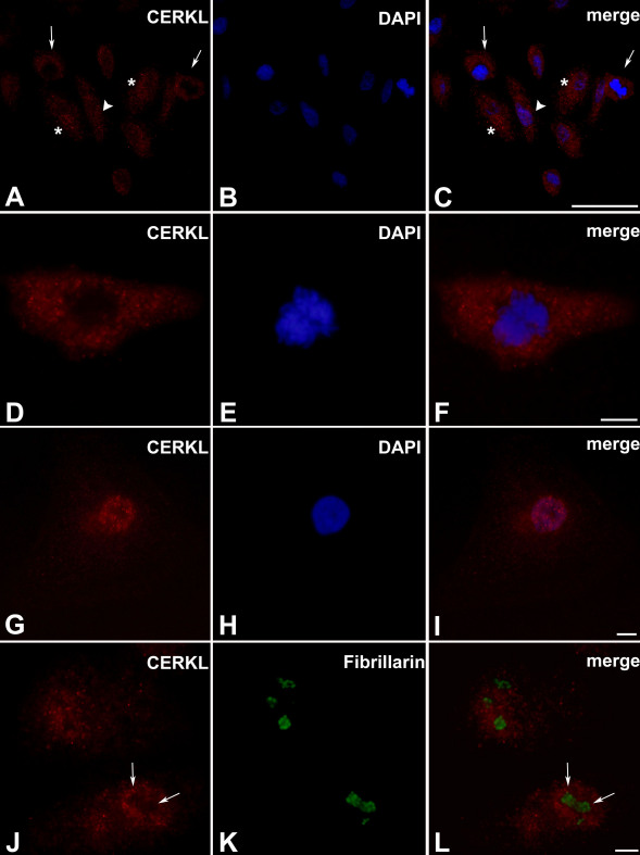
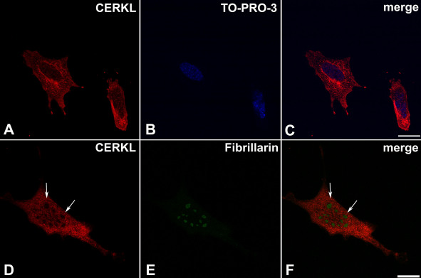
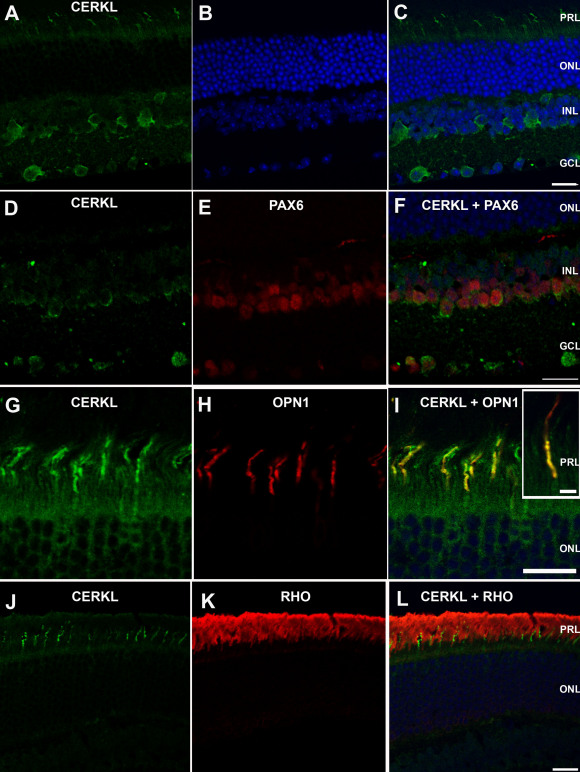
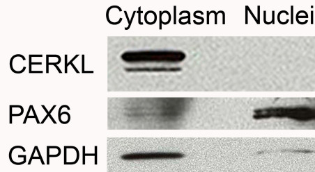
Results: Cerkl is expressed in the mouse eye as early as embryonic day 14. A total of seven different Cerkl splice-isoforms were identified in the mouse retina. The subcellular localization of CERKL in retina-derived cell lines is variable: CERKL is diffusely distributed in the cytoplasm, and in many cells, it is highly concentrated in the perinuclear region. In most, but not all cells, CERKL is also highly concentrated in the nucleus. In the mouse retina, CERKL is located in the ganglion cell layer, in amacrine cells of the inner nuclear layer, and in photoreceptors. CERKL is highly expressed in cone photoreceptors; however, its expression level in rod photoreceptors is very low. In cultured cells, CERKL is detected in the nucleus, but in retinal cells in situ, it is mostly located in the cytoplasm.

Conclusions: The expression of Cerkl in both mature and embryonic mouse retina and the severe retinal phenotype associated with human CERKL mutations indicate that this gene plays a crucial role in retinal activity, and that it may be important for retinal development as well. The high expression level of CERKL in cones correlates with the CERKL-associated phenotype in humans. Whether nucleocytoplasmic transport of CERKL actually occurs in vivo under certain conditions and its functional significance remain to be discovered.


Figures

Figure 1
Figure 1
Expression analysis of Cerkl in the mouse eye. A: Reverse Transcription-Polymerase Chain Reaction (RT–PCR) analysis of Cerk (412 bp) and Cerkl expression in mouse brain (Br) and retina (Ret). Cerkl expression was tested using primers located in exons 1 and 2 (see Figure 2B). The two PCR products obtained for Cerkl (503 and 363 bp) represent different splice-isoforms. B: RT–PCR analysis of Cerkl in the mouse eye at different developmental time points: embryonic day 14 (E14), newborn (P0), and 5 months (5 mo). The analysis indicates Cerkl expression at all time points tested. β-actin (Actb; 437 bp product) served as an internal control for RNA quality and quantity. M: size marker.
Figure 2
Figure 2
CERKL gene and splice-variants in human and mouse retina. A schematic representation of CERKL genes (drawn to scale), splice-variants (left panels), and expected protein products (right panels) is shown. In the splice-variants illustrations, filled boxes represent coding exons, and open boxes represent non-coding exons. Alternatively spliced exons are marked in red. In the protein products illustrations, the PH, DAGK, and CERK homology domains are indicated. The borders of each domain are marked by their amino acid positions, indicated above each protein. The breaks between exons are indicated below each protein. Protein molecular weight in kDa is shown on the right. A: Human CERKL gene and splice-variants previously identified in the human retina. Variants e and f correspond to AY690333 and AY690332 [13]. B: Mouse Cerkl gene and splice-variants found in the mature and embryonic (E14) mouse retina. Two of the mouse isoforms (isoforms d’ and g’) encode for the same short protein product. Mouse isoform a’, which is present in both adult and embryonic samples, is equivalent to human isoform a, and mouse isoform f’ is equivalent to human isoform f. Locations of PCR primers used for RT–PCR analysis and for splice-variant identification (forward primer located in exon 1 and reverse primers located in exons 2 and 14) are indicated by arrows above the schematic representation of the murine gene.
Figure 3
Figure 3
Verification of the specificity of the anti-CERKL antibody RA. A: Western blot analysis of mouse retinal extract with the affinity-purified RA antibody. The antibody detects a main specific band, which corresponds to the expected size of the primary and most abundant CERKL isoform in the adult mouse retina (isoform a’, 58 kDa). Two additional fainter bands slightly higher than 51 kDa, corresponding to isoforms b’ and c’ (55 and 53 kDa, respectively), can also be observed. All three bands are completely absent following pre-absorption of the RA antibody with the recombinant CERKL protein (right panel), but not with a non-specific protein (BSA; left panel). B: Western blot analysis of extracts from bacteria transformed with mouse Cerkl retinal isoforms a’ (58 kDa), b’ (55 kDa), and d’ (12 kDa). The RA antibody detects proteins of the expected sizes in IPTG-induced, but not in un-induced bacterial extracts. C: Western blot analysis of protein extracts from the ARPE-19 and 661W cell lines. In the mouse-derived cell line, 661W, a major band of approximately 58 kDa, which corresponds to mouse CERKL isoform a’ is detected. In the human-derived cell line, ARPE-19, two bands corresponding to human CERKL isoforms c and d (46 and 51 kDa, respectively) are detected. D: Immunostaining of ARPE-19 cells is omitted following pre-absorption of the RA antibody (green) with the recombinant CERKL protein (right panel), but not with a non-specific protein (BSA; left panel). Nuclei are stained with TO-PRO-3 (blue). Scale bar, 20 µm. E: Immunostaining of a mouse retina section is omitted following pre-absorption of the RA antibody (green) with the recombinant CERKL protein (right panel), but not with a non-specific protein (BSA; left panel). Nuclei are stained with TO-PRO-3 (blue). Scale bar, 20 µm.
Figure 4
Figure 4
CERKL subcellular localization in ARPE-19 cells. Cells were stained with the RA anti-CERKL antibody (red) and with DAPI for nuclear staining (blue). A-C: The subcellular localization of CERKL is variable. It can be located in both the nucleus and the cytoplasm (arrowhead), or only in the cytoplasm (arrows). In many cells, CERKL is concentrated in the perinuclear region (asterisks). Scale bar, 50 µm. D-F: A cell in which CERKL is absent from the nucleus. Scale bar, 10 µm. G-I: A cell in which CERKL is concentrated in the nucleus. Scale bar, 10 µm. J-L: CERKL is absent from nucleoli (arrows), which are stained by the nucleolar marker Fibrillarin (green). Scale bar, 10 µm.
Figure 5
Figure 5
CERKL subcellular localization in 661W cells. Cells were stained with the RA anti-CERKL antibody (red) and with TO-PRO-3 for nuclear staining (blue). A-C: CERKL can be located in both the nucleus and the cytoplasm, where it is concentrated in the perinuclear region. Scale bar, 20 µm. D-F: CERKL is absent from nucleoli (arrows), which are stained by the nucleolar marker Fibrillarin (green). Scale bar, 20 µm.
Figure 6
Figure 6
CERKL expression pattern in the mouse retina. Serial sagittal sections of adult mouse retina were immunostained with the RA anti-CERKL antibody. A-C: CERKL staining (green) shows expression in the photoreceptor cell layer (PRL), the inner nuclear layer (INL), and the ganglion cell layer (GCL). D-F: Double staining for CERKL (green) and PAX6 (red), a marker for amacrine cells, in the INL and GCL. G-I: Double staining for CERKL (green) and OPN1 (red), a marker for cone photoreceptor cells. The insert in panel I shows a higher magnification of a double-stained cone photoreceptor. J-L: Double staining for CERKL (green) and RHO (rhodopsin; red), a marker for rod photoreceptor cells. Nuclei are stained with TO-PRO-3 (blue). Note that CERKL is highly expressed in cone photoreceptors, while its expression level in rod photoreceptors is very low. No staining was observed when sections were stained with serum from pre-immune rabbits or with secondary antibody only (data not shown). PRL, photoreceptor layer; ONL, outer nuclear layer; INL, inner nuclear layer; GCL, ganglion cell layer. Scale bars, 20 µm.
Figure 7
Figure 7
CERKL expression in the cytoplasm and nuclei of adult mouse retina. Western blot analysis was conducted on nuclear and cytoplasmic fractions of adult mouse retinas with an anti-CERKL antibody. PAX6 served as a positive control for the nuclear fraction. GAPDH served as a positive control for the cytoplasmic fraction.

References

    1. Rivolta C, Sharon D, DeAngelis MM, Dryja TP. Retinitis pigmentosa and allied diseases: numerous diseases, genes, and inheritance patterns. Hum Mol Genet. 2002;11:1219–27. - PubMed
    1. Hamel CP. Cone rod dystrophies. Orphanet J Rare Dis. 2007;2:7. - PMC - PubMed
    1. Hartong DT, Berson EL, Dryja TP. Retinitis pigmentosa. Lancet. 2006;368:1795–809. - PubMed
    1. Bayés M, Goldaracena B, Martinez-Mir A, Iragui-Madoz MI, Solans T, Chivelet P, Bussaglia E, Ramos-Arroyo MA, Baiget M, Vilageliu L, Balcells S, Gonzalez-Duarte R, Grinberg D. A new autosomal recessive retinitis pigmentosa locus maps on chromosome 2q31-q33. J Med Genet. 1998;35:141–5. - PMC - PubMed
    1. Tuson M, Marfany G, Gonzalez-Duarte R. Mutation of CERKL, a novel human ceramide kinase gene, causes autosomal recessive retinitis pigmentosa (RP26). Am J Hum Genet. 2004;74:128–38. - PMC - PubMed

Publication types

MeSH terms

Substances