

Late onset of cerebellar abiotrophy in a boxer dog
- PMID: 21151662
- PMCID: PMC2997505
- DOI: 10.4061/2010/406275
Late onset of cerebellar abiotrophy in a boxer dog
Abstract
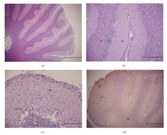
Cerebellar abiotrophy is a degenerative disorder of the central nervous system and has been reported in humans and animals. This case report documents clinical, histopathological, and immunohistochemical findings of cerebellar abiotrophy in an adult Boxer dog. A 3.5-year-old, female, tan Boxer dog presented with a six-week history of left-sided head tilt. Neurological examination and additional diagnostics during her three subsequent visits over 4.5 months revealed worsening of neurological signs including marked head pressing, severe proprioceptive deficits in all the four limbs, loss of menace response and palpebral reflex in the left eye, and a gradual seizure lasting one hour at her last visit. Based on the immunohistochemical staining for glial fibrillary acidic protein and histopathological examination of cerebellum, cerebellar cortical abiotrophy was diagnosed. This is the first reported case of cerebellar abiotrophy in a Boxer dog to our knowledge.
Figures
References
-
- Summers BA, Cummings JF, De Lahunta A. Degenerative diseases of the central nervous system. In: Summers BA, Cummings JF, De Lahunta A, editors. Veterinary Neuropathology. St. Louis, Mo, USA: Mosby-Year Book; 1995. pp. 300–305.
-
- Mouser P, Lévy M, Sojka JE, Ramos-Vara JA. Cerebellar abiotrophy in an alpaca (Lama pacos) Veterinary Pathology. 2009;46(6):1133–1137. - PubMed
-
- De Lahunta A, Glass E. Cerebellum. In: De Lahunta A, Glass E, editors. Veterinary Neuroanatomy and Clinical Neurology. St. Louis, Mo, USA: Elsevier; 2009. pp. 343–388.
-
- Hartley WJ, Barker JSF, Wanner RA, Farrow BR. Inherited cerebellar degneration in the rough coated collie. Australian Veterinary Practitioner. 1978;8:79–85.
Publication types
LinkOut - more resources
Full Text Sources
