. 2010:2010:124509.
doi: 10.1155/2010/124509. Epub 2010 Dec 1.

Importance of Lipopolysaccharide and Cyclic β-1,2-Glucans in Brucella-Mammalian Infections

Affiliations

Importance of Lipopolysaccharide and Cyclic β-1,2-Glucans in Brucella-Mammalian Infections

Andreas F Haag et al. Int J Microbiol. 2010.

Abstract

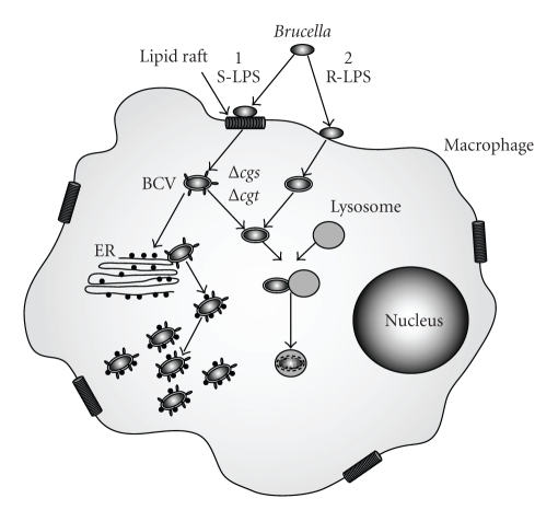
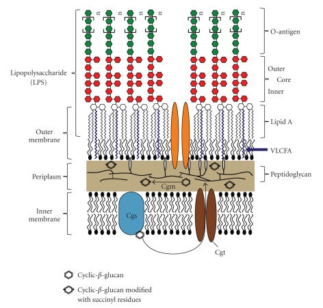
Brucella species are the causative agents of one of the most prevalent zoonotic diseases: brucellosis. Infections by Brucella species cause major economic losses in agriculture, leading to abortions in infected animals and resulting in a severe, although rarely lethal, debilitating disease in humans. Brucella species persist as intracellular pathogens that manage to effectively evade recognition by the host's immune system. Sugar-modified components in the Brucella cell envelope play an important role in their host interaction. Brucella lipopolysaccharide (LPS), unlike Escherichia coli LPS, does not trigger the host's innate immune system. Brucella produces cyclic β-1,2-glucans, which are important for targeting them to their replicative niche in the endoplasmic reticulum within the host cell. This paper will focus on the role of LPS and cyclic β-1,2-glucans in Brucella-mammalian infections and discuss the use of mutants, within the biosynthesis pathway of these cell envelope structures, in vaccine development.


Figures

Figure 1
Figure 1
The Brucella-macrophage interaction. The preferred cells infected by Brucella are macrophages. Brucella strains with smooth LPS (S-LPS) enter the cell through interaction with lipid rafts and are then encompassed in a membrane bound compartment called Brucella containing vacuole (BCV). This vacuole retains some lipid raft markers, targeting the BCV to the endoplasmic reticulum (ER). Brucella fuses with the ER, thus acquiring ER markers to avoid fusion with the lysosome before beginning to replicate. Rough LPS mutants do not enter the macrophage through lipid rafts and are rapidly targeted to the lysosome and killed. Mutants in the CβG biosynthesis pathway (Δcgs and Δcgt) do not fuse with the ER but are targeted to the lysosome.
Figure 2
Figure 2
Diagrammatic representation of the Brucella cell envelope. The cell envelope is comprised of an inner membrane, consisting of a bilayer of phospholipids, and an outer membrane with an inner leaflet of phospholipids and an outer leaflet of lipopolysaccharide (LPS). LPS consists of three components. The O-antigen faces the extracellular space and it is the component that is recognized by the adaptive immune response. The O-antigen is connected to a sugar core molecule composed of different sugars which have not yet been fully identified. Lipid A forms the hydrophobic anchor of LPS within the membrane and has a backbone of diaminoglucose, which is acylated with saturated and hydroxylated fatty acids. Brucella lipid A contains an unusual very-long-chain fatty acid (VLCFA). Cyclic β-1,2-glucans are synthesized by the inner membrane protein Cgs and then transported to the periplasm by the predicted ABC-transporter Cgt where they are modified with on average two succinyl residues by a predicted membrane protein Cgm.
Figure 3
Figure 3
Genomic organization of O-antigen and core biosynthesis genes. The genes predicted to be involved in the biosynthesis of B. melitensis LPS O-antigen and core molecule are distributed at four different loci throughout the genome. The majority of the genes are located in the wbk region and the wbo region on chromosome I. Genes involved in the core biosynthesis can be found on chromosome I (w a**) and chromosome II (m a n B core and m a n C core). The wbk region contains multiple insertion sequences (ISs) suggesting that this region was acquired through horizontal gene transfer [22].
Figure 4
Figure 4
Predicted pathways of LPS O-antigen and core molecule biosynthesis. Brucellae derive all sugars from glucose. Green arrows indicate the reactions leading to the synthesis of the monomeric O-antigen subunits, which are polymerized onto bactoprenol (pathway indicated in blue). The complete O-antigen is then transported to the periplasmic face of the inner membrane by the ABC transporter system Wzm/Wzt and ligated to lipid A molecule by the O-antigen ligase WaaL. Pathways leading to the biosynthesis of the core molecule are indicated in red. Hexagons indicate whether mutants in the respective genes are affected in the biosynthesis of the O-antigen (OAg) or the sugar core molecule (Core). Dotted arrows indicate abbreviated pathways involving the defined enzymes.

References

    1. Pappas G, Papadimitriou P, Akritidis N, Christou L, Tsianos EV. The new global map of human brucellosis. Lancet Infectious Diseases. 2006;6(2):91–99. - PubMed
    1. Young EJ. An overview of human brucellosis. Clinical Infectious Diseases. 1995;21(2):283–290. - PubMed
    1. Meyer KF, Shaw EB. A comparison of the morphologic, cultural and biochemical characteristics of B. abortus and B. melitensis from cattle. Studies on the genus Brucella nov. gen. Journal of Infectious Diseases. 1920;27:173–184.
    1. Huddleson IF. Differentiation of the species of the genus Brucella . American Journal of Public Health. 1931;21(5):491–498. - PMC - PubMed
    1. Carmichael LE, Bruner DW. Characteristics of a newly-recognized species of Brucella responsible for infectious canine abortions. The Cornell Veterinarian. 1968;48(4):579–592. - PubMed

LinkOut - more resources