. 2010 Nov 30;1(5):e00265-10.
doi: 10.1128/mBio.00265-10.

Rotavirus disrupts calcium homeostasis by NSP4 viroporin activity

Affiliations

Rotavirus disrupts calcium homeostasis by NSP4 viroporin activity

Joseph M Hyser et al. mBio. .

Abstract

Many viruses alter intracellular calcium homeostasis. The rotavirus nonstructural protein 4 (NSP4), an endoplasmic reticulum (ER) transmembrane glycoprotein, increases intracellular levels of cytoplasmic Ca(2+) ([Ca(2+)]cyto) through a phospholipase C-independent pathway, which is required for virus replication and morphogenesis. However, the NSP4 domain and mechanism that increases [Ca(2+)]cyto are unknown. We identified an NSP4 domain (amino acids [aa] 47 to 90) that inserts into membranes and has structural characteristics of viroporins, a class of small hydrophobic viral proteins that disrupt membrane integrity and ion homeostasis to facilitate virus entry, assembly, or release. Mutational analysis showed that NSP4 viroporin activity was mediated by an amphipathic α-helical domain downstream of a conserved lysine cluster. The lysine cluster directed integral membrane insertion of the viroporin domain and was critical for viroporin activity. In epithelial cells, expression of wild-type NSP4 increased the levels of free cytoplasmic Ca(2+) by 3.7-fold, but NSP4 viroporin mutants maintained low levels of [Ca(2+)]cyto, were retained in the ER, and failed to form cytoplasmic vesicular structures, called puncta, which surround viral replication and assembly sites in rotavirus-infected cells. When [Ca(2+)]cyto was increased pharmacologically with thapsigargin, viroporin mutants formed puncta, showing that elevation of calcium levels and puncta formation are distinct functions of NSP4 and indicating that NSP4 directly or indirectly responds to elevated cytoplasmic calcium levels. NSP4 viroporin activity establishes the mechanism for NSP4-mediated elevation of [Ca(2+)]cyto, a critical event that regulates rotavirus replication and virion assembly.


Figures

FIG 1
FIG 1
The amphipathic domain mediates membrane permeabilization. (A) Linear schematic of NSP4 and the primary sequence of the viroporin domain highlighting the five conserved lysines (blue) and two conserved cysteines (arrowheads). H1 and H2, hydrophobic domains 1 and 2, respectively; CCD, coiled-coil domain; DLP-R, double-layered particle receptor domain. Helical wheel representations of the pentalysine domain (PD) and amphipathic domain (AD). Black, hydrophobic residues; green, polar uncharged residues; blue, basic residues; red, acidic residues. (B) Schematic of the NSP4 deletions tested. The membrane-destabilizing activity (MDA) is summarized at the right (+, activity; −, no activity). (C) The OD600 of uninduced (−IPTG, black line) or NSP4-expressing cultures were determined at 10-min intervals for 90 minutes and presented as the percent optical density relative to the optical density at the time of induction. (D) Immunoblot analysis of NSP4 expression. Fivefold more lysate was loaded in the bottom immunoblot to detect NSP447-90. (E) Oligomerization of partially purified NSP447-90 was analyzed by immunoblot analysis after SDS-PAGE in the absence or presence of β-mercaptoethanol. m, monomer; d, dimer; o, oligomer.
FIG 2
FIG 2
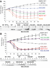
Positive charges of amino acids 62, 66, and 69 are crucial for NSP4 viroporin activity. (A, top) Lysine-to-glutamic acid mutants were tested in the E. coli lysis assay. The OD600 of uninduced (−IPTG, black line) or NSP4-expressing cultures was determined at 10-min intervals for 90 minutes and presented as the percent optical density relative to the optical density at the time of induction. (Bottom) Immunoblot of NSP4 expression using monoclonal antibody B4-2/55. (B, top) Lysine-to-alanine and lysine-to-histidine mutants were tested in the E. coli lysis assay, as described above. (Bottom) Immunoblot of NSP4 expression using monoclonal antibody B4-2/55.
FIG 3
FIG 3
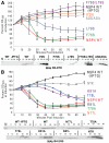
The amphipathic α-helix is crucial for NSP4 viroporin activity. (A) Mutations of the nonpolar surface of the amphipathic domain were tested in the E. coli lysis assay. (Top) The OD600 of uninduced (−IPTG, black line) or NSP4-expressing cultures was determined at 10-min intervals for 90 minutes and presented as the percent optical density relative to the optical density at the time of induction. (Bottom) Immunoblot of NSP4 expression using monoclonal antibody B4-2/55. (B, top) Single and multiple mutations of the polar surface of the amphipathic domain were tested in the E. coli lysis assay, as described above. (Bottom) Immunoblot of NSP4 expression using monoclonal antibody B4-2/55.
FIG 4
FIG 4
NSP4 cytotoxicity is mediated by the viroporin domain. Serial dilutions of the indicated constructs were plated on plates with LB-Amp and LB-Amp plus IPTG and incubated overnight at 37°C. The difference in the numbers of CFU/milliliter between LB-Amp and LB-Amp plus IPTG is graphed. *, P < 0.01.
FIG 5
FIG 5
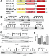
The pentalysine motif mediates integral membrane insertion of the viroporin domain. (A) Schematic of the NSP4 constructs tested. (B and C) Immunoblot analysis of NSP4 in total cell lysate (T), soluble protein (S), peripheral membrane protein (P), and integral membrane protein (I) fractions. M, molecular weight marker. (D) Immunoblot analysis of MA104 cell lysates for WT NSP4-EGFP, K62,66,69E, and ASDASA in the absence or presence of MG132. Lysates were mock treated or Endo H treated to demonstrate the glycosylation of NSP4 (gly) by shifting to the unglycosylated form (ungly). (E) Flow cytometry analysis of the mean fluorescence intensity (MFI) of MA104 cells expressing WT NSP4-EGFP or K62,66,69E in the absence (dark gray) or presence (light gray) of MG132. *, P < 0.01 for K62,66,69E versus WT in the absence of MG132; #, P < 0.01 for K62,66,69E absence versus the presence of MG132. AU, arbitrary units. (F) Immunoblot analysis of MA104 fractionation, as described above, for WT, K62,66,69E, and ASDASA.
FIG 6
FIG 6
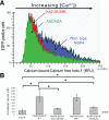
NSP4 viroporin mutants do not elevate cytoplasmic calcium levels. (A) HEK293T cells expressing EGFP, WT NSP4-EGFP, or the indicated viroporin mutant NSP4-EGFP were loaded with 1.8 µM Indo-1 and analyzed by flow cytometry to measure the levels of cytoplasmic calcium. RFU, relative fluorescence units. (B) The calcium-bound/calcium-free Indo-1 ratio (R) was determined for 10,000 EGFP-positive cells, and the [Ca2+]cyto was calculated. A total of 3 independent experiments were performed, and error bars indicate the standard deviations of the means. *, P < 0.05.
FIG 7
FIG 7
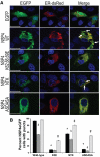
Mutation of the viroporin domain blocks spontaneous NSP4-EGFP puncta formation but not the ability to form puncta after Ca2+ stimulation. (A) Confocal microscopy images of cells expressing EGFP, WT NSP4-EGFP, or viroporin mutants (first column), the DsRed-ER marker of the endoplasmic reticulum compartment (second column), and the merged images (third column). White arrows indicate the characteristic NSP4-EGFP punctate structures. Bar = 20 µm. (B) NSP4-EGFP-expressing cells were scored for punctate or reticular EGFP signal in normal medium (black), a 50-µM BAPTA-AM treatment (dark grey), or a 1-µM TG treatment (light grey). *, P < 0.01 for the mutant versus the WT in normal media. &, P < 0.01 for the mutant in BAPTA versus in normal medium; #, P < 0.01 for the mutant in TG versus in normal medium.
FIG 8
FIG 8
Model of the NSP4 viroporin as a three-pass transmembrane protein. (Left) Initial insertion of NSP4 into the ER membrane (gray) occurs through the uncleaved signal sequence in the H2 domain. Lysine residues interact with ER membrane phospholipids and promote insertion of the viroporin domain as an anti-parallel α-helical hairpin. (Center) Insertion of the viroporin domain generates a three-pass transmembrane topology. (Right) Oligomerization of NSP4 around the amphipathic α-helix creates an aqueous pore through the membrane and allows the release of ER Ca2+.

Similar articles

Cited by

References

    1. Dubyak G. R. 2004. Ion homeostasis, channels, and transporters: an update on cellular mechanisms. Adv. Physiol. Educ. 28:143–154 - PubMed
    1. Kang M., Moroni A., Gazzarrini S., DiFrancesco D., Thiel G., Severino M., Van Etten J. L. 2004. Small potassium ion channel proteins encoded by chlorella viruses. Proc. Natl. Acad. Sci. U. S. A. 101:5318–5324 - PMC - PubMed
    1. Daniels R., Sadowicz D., Hebert D. N. 2007. A very late viral protein triggers the lytic release of SV40. PLoS Pathog. 3:e98 - PMC - PubMed
    1. Suzuki T., Orba Y., Okada Y., Sunden Y., Kimura T., Tanaka S., Nagashima K., Hall W. W., Sawa H. 2010. The human polyoma JC virus agnoprotein acts as a viroporin. PLoS Pathog. 6:e1000801 - PMC - PubMed
    1. Liao Y., Tam J. P., Liu D. X. 2006. Viroporin activity of SARS-CoV E protein. Adv. Exp. Med. Biol. 581:199–202 - PMC - PubMed

Publication types

LinkOut - more resources