. 2010 Dec 8:4:202.
doi: 10.3389/fnins.2010.00202. eCollection 2010.

Long-Range GABAergic Connections Distributed throughout the Neocortex and their Possible Function

Affiliations

Long-Range GABAergic Connections Distributed throughout the Neocortex and their Possible Function

Nobuaki Tamamaki et al. Front Neurosci. .

Abstract

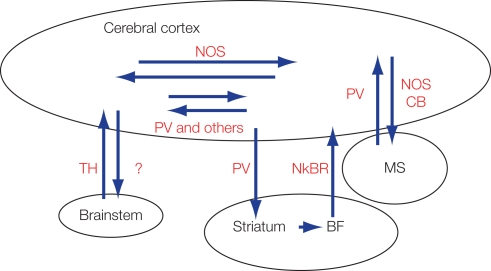
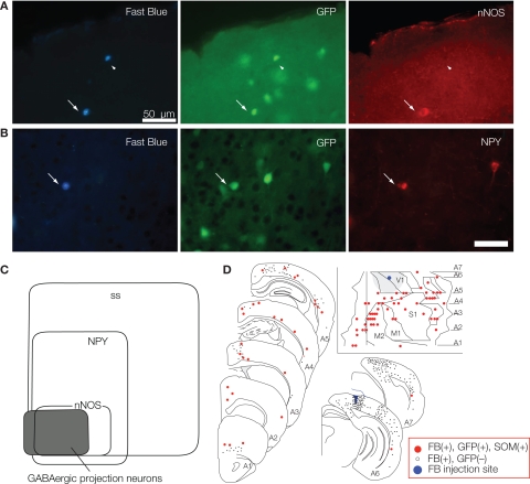
Features and functions of long-range GABAergic projection neurons in the developing cerebral cortex have been reported previously, although until now their significance in the adult cerebral cortex has remained uncertain. The septo-hippocampal circuit is one exception - in this system, long-range mature GABAergic projection neurons have been well analyzed and their contribution to the generation of theta-oscillatory behavior in the hippocampus has been documented. To have a clue to the function of the GABAergic projection neurons in the neocortex, we view how the long-range GABAergic projections are integrated in the cortico-cortical, cortico-fugal, and afferent projections in the cerebral cortex. Then, we consider the possibility that the GABAergic projection neurons are involved in the generation, modification, and/or synchronization of oscillations in mature neocortical neuron activity. When markers that identify the GABAergic projection neurons are examined in anatomical and developmental studies, it is clear that neuronal NO synthetase (nNOS)-immunoreactivity can readily identify GABAergic projection neurons. GABAergic projection neurons account for 0.5% of the neocortical GABAergic neurons. To elucidate the role of the GABAergic projection neurons in the neocortex, it will be necessary to clarify the network constructed by nNOS-positive GABAergic projection neurons and their postsynaptic targets. Thus, our long-range goals will be to label and manipulate (including deleting) the GABAergic projection neurons using genetic tools driven by a nNOS promoter. We recognize that this may be a complex endeavor, as most excitatory neurons in the murine neocortex express nNOS transiently. Nevertheless, additional studies characterizing long-range GABAergic projection neurons will have great value to the overall understanding of mature cortical function.

Keywords: GABA; GABAergic projection neuron; gamma-oscillations; nNOS; neocortex.


Figures

Figure 1
Figure 1
Currently reported cortico-cortical, cortico-fugal, and afferent GABAergic projections. Most of these GABAergic projection neurons express characteristic protein or peptides. BF, basal forebrain; CB, calbindin; MS, medial septum; NkBR, neurokinin-B receptor; nNOS, neuronal NO synthetase; PV, parvalbumin; SS, somatostatin; TH, tyrosine hydroxylase.
Figure 2
Figure 2
Following FB injection into a visual neocortical area of the GAD67-GFP mouse, we found temporal neocortical areas that contained FB- and GFP-double-labeled cells but few FB-single-labeled cells. Most of these double-labeled neurons showed immunoreactivity of somatostatin, neuropeptide Y (A), and nNOS (B). A Venn diagram in (C) shows the relationship between the immunoreactivity and the GABAergic projection neurons in the neocortex. (D) Distribution of SS-positive GABAergic projection neurons were revealed by FB injection into the neocortex V1 and represented on the mouse-flattened cortical map and serial frontal sections. Each small gray filled circle represents 20 FB-single-labeled neurons. Each red-filled circle indicates an FB-, GFP- and SS-positive neuron. Blue marker and gray large circle (insets) shows the FB injection site and cortical region within 1.5 mm from the FB injection site, respectively. Calibration bar in (B) is 50 μm. (C,D) Adapted from Tomioka et al. (2005).
Figure 3
Figure 3
Synchrony of gamma-oscillation in cat visual cortices. Callosal connections mediate long-distance synchronization. In the bottom panels are cross-correlograms between responses from different recording sites in the left hemisphere (LH) and right hemisphere (RH) that indicate the degree of interhemispheric synchronization. When the corpus callosum was intact (left-hand panel), strong interhemispheric synchronization occurred with no phase lag between the LH and RH recording sites. Sectioning of the corpus callosum (right-hand panel) abolished interhemispheric synchronization while leaving synchronization within hemispheres intact. Adapted from Uhlhaas and Singer (2010).
Figure 4
Figure 4
GABAergic subplate neurons in the intermediate zone (IZ) also extend their axon cortico-fugally. (A) Postnatal day 0 rat neocortex. Top and second arrows indicate the width of subplate according to Bayer and Altman (1991). Second and third arrows indicate the width of white matter which contains many GABAergic neurons. Double-arrows indicate the area where we found retrogradely labeled neurons after lipophilic red fluorescent dye, DiI injection into the median plane of the spinal cord. See insets in (B). (B) DiI injection into the spinal cord (C1–C3) at P0 labeled pyramidal neurons in layer V and subplate neurons in the IZ. Axons of pyramidal neurons followed those of GABAergic subplate neurons rather than those of GABA-negative subplate neurons (layer VII). (C) Biotin-dextan-amine (BDA) injection into the primordium of pons at E18 rat labeled subplate neurons in the IZ at P0. CP, cortical plate; IZ, intermediate zone; S, subplate containing excitatory subplate neurons. Arrow heads in the insets in (B,C) indicate the injection site of DiI and BDA. Calibration bars in (A), and in the insets in (B,C) are 1 mm. Calibration bar in (B) is 100 μm, that in (C) is 50 μm. (B) Adapted from Tamamaki et al. (1997a,b).

References

    1. Albus K., Wahle P. (1994). The topography of tangential inhibitory connections in the postnatally developing and mature striate cortex of the cat. Eur. J. Neurosci. 6, 779–79210.1111/j.1460-9568.1994.tb00989.x - DOI - PubMed
    1. Anderson S. A., Eisenstat D. D., Shi L., Rubenstein J. L. (1997). Interneuron migration from basal forebrain to neocortex: dependence on Dlx genes. Science 278, 474–47610.1126/science.278.5337.474 - DOI - PubMed
    1. Aroniadou-Anderjaska V., Keller A. (1996). Intrinsic inhibitory pathways in mouse barrel cortex. Neuroreport 7, 2363–236810.1097/00001756-199610020-00017 - DOI - PubMed
    1. Bayer S. A., Altman J. (1991). Neocortical Development. New York: Raven Press
    1. Bielle F., Griveau A., Narboux-Nême N., Vigneau S., Sigrist M., Arber S., Wassef M., Pierani A. (2005). Multiple origins of Cajal–Retzius cells at the borders of the developing pallium. Nat. Neurosci. 8, 1002–101210.1038/nn1511 - DOI - PubMed

LinkOut - more resources