

doi: 10.1557/mrs2010.571.
Shape and Biomechanical Characteristics of Human Red Blood Cells in Health and Disease
- PMID: 21151848
- PMCID: PMC2998922
- DOI: 10.1557/mrs2010.571
Item in Clipboard
Shape and Biomechanical Characteristics of Human Red Blood Cells in Health and Disease
MRS Bull.
2010 May.
Abstract
The biconcave shape and corresponding deformability of the human red blood cell (RBC) is an essential feature of its biological function. This feature of RBCs can be critically affected by genetic or acquired pathological conditions. In this review, we highlight new dynamic in vitro assays that explore various hereditary blood disorders and parasitic infectious diseases that cause disruption of RBC morphology and mechanics. In particular, recent advances in high-throughput microfluidic devices make it possible to sort/identify healthy and pathological human RBCs with different mechanobiological characteristics.
Figures
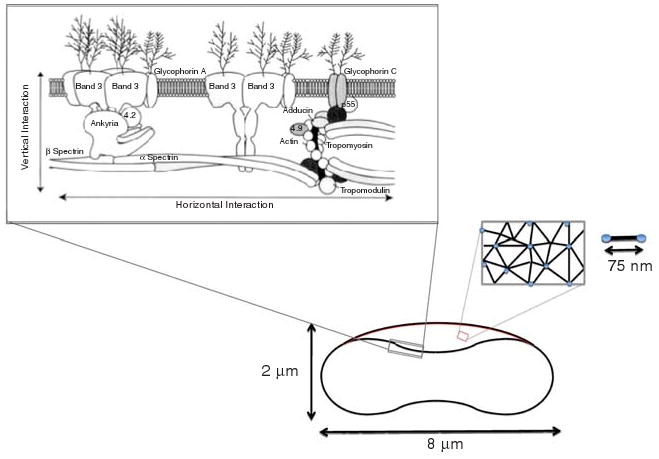
Schematic representation of a healthy red blood cell (RBC) membrane, geometry (discocyte shape), and spectrin network. Upper left inset shows the cross-sectional view of the RBC membrane with vertical and horizontal interactions. Right inset shows the two-dimensional connectivity of the spectrin network. Reprinted with permission from Reference 2. ©1999, Wiley.
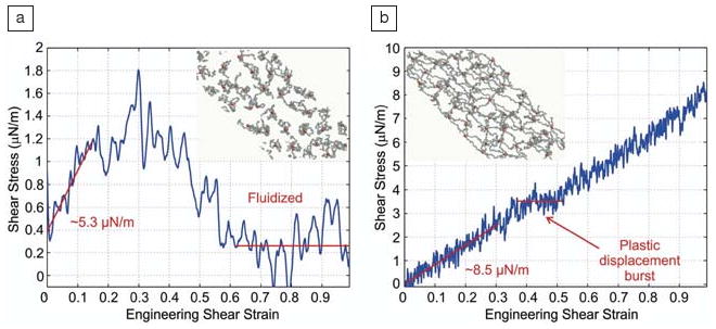
Shear stress-strain response of a spectrin network at a strain rate of 3 × 10⁵ per s. (a) Energy hit rate 10 per s and final network structure (inset) at 100% shear strain. (b) Energy hit rate 2.5 per s and final network structure (inset) at 100% shear strain. Reprinted with permission from Reference 9. ©2007, National Academy of Sciences.
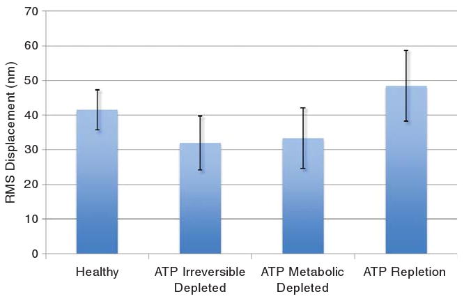
Effect of adenosine-5′-triphosphate (ATP) on membrane fluctuations measured by root mean squared (RMS) displacement of red blood cell membrane. When ATP is depleted, irreversibly or metabolically, membrane fluctuations decrease, and echinocyte shape is observed. When ATP levels are restored, normal fluctuation behavior and biconcave shape returns. Adapted from Reference 12.
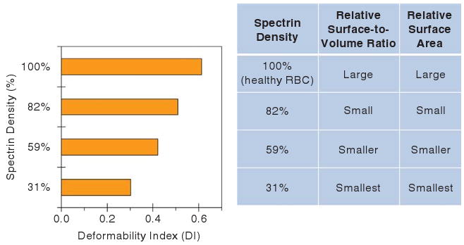
Deformability of the healthy and spherocyte red blood cells (RBCs) as a function of spectrin concentration at a fixed osmolality of 300 mOs mol/kg. The lower the spectrin density, the smaller the deformability index (DI) values, surface-to-volume ratio, and surface area. In more hypotonic solutions (lower osmolality), a decrease in surface-to-volume ratio causes a reduction in DI at a fixed spectrin density. In hypertonic solutions (higher osmolality), cellular dehydration results in lower DI. Adapted from Reference 19.
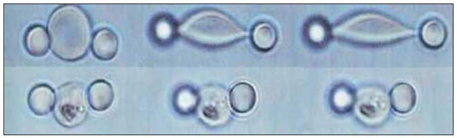
Controlled stretching using an optical trap of healthy (top) and infected (bottom) red blood cells (RBCs). The left lighted bead indicates that it was trapped by the laser, while the right bead was adhered to the glass cover slip. Bottom row shows how stiff the late-stage P. falciparum–infected RBC (Pf-RBCs) had become when compared with its healthy counterpart shown in the top row undergoing the same stretching force., (Bead diameter is 4 μm.) Reprinted with permission from Reference 34. ©2005, Elsevier.
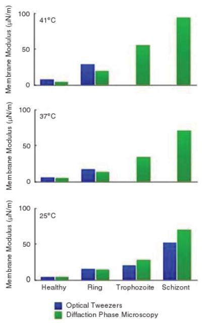
Shows the shear modulus response of infected red blood cell (RBC) membrane as a function of the P. falciparum intra-erythrocytic developmental cycle (ring, trophozoite, and schizont stages) and temperature. Data from two independent experimental techniques, optical tweezers and diffraction phase microscopy, show good agreement.,,

Passage of (a) uninfected, (b) trophozoite, and schizont stages of P. falciparum–infected red blood cells (RBCs) (Pf-RBCs) and (c–e) ring, trophozoite, and schizont stages of P. vivax–infected RBCs (Pv-RBCs) through 2 μm constricted channels. Scale bar = 10 μm in all cases. Reprinted with permission from Reference 42. ©2009, University of Chicago Press.
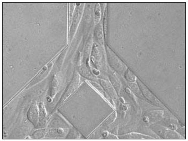
A bifurcated micochannel showing cyto-adhered P. falciparum infected RBC (Pf-RBCs). The microchannel has been seeded with Chinese Hamster Ovary (CHO) cells expressing CD-36 human receptor. (Pf-RBCs average diameter of 7.5 μm). Reprinted with permission from Reference 55. ©2008, Wiley.
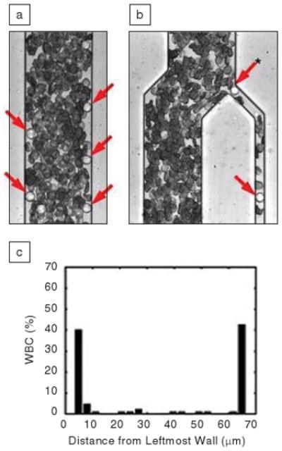
Microfluidic blood cell sorting using the Fahraeus effect. (a) In a 5.5-mm long rectangular, straight microchannel leukocytes will be marginated to the edge, while more flexible red blood cells (RBCs) are concentrated in the center. (b) Using a bifuricated channel, one can sample and enrich white blood cells (WBCs), which are indicated by arrows. (c) The separation process is determined by both size and deformability of the WBC, compared with RBC. Reprinted with permission from Reference 60. ©2005, American Chemical Society.
References
Grants and funding
LinkOut - more resources
Full Text Sources
Other Literature Sources