

Xanthomatous infiltration of the rotator cuff and long head of biceps with rotator cuff tear in a patient with mixed hyperlipidemia: a case report with MRI imaging
- PMID: 21151852
- PMCID: PMC2998979
- DOI: 10.4137/CMAMD.S3172
Xanthomatous infiltration of the rotator cuff and long head of biceps with rotator cuff tear in a patient with mixed hyperlipidemia: a case report with MRI imaging
Abstract
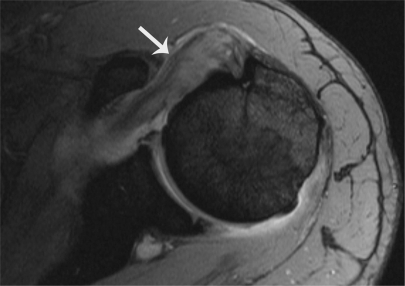
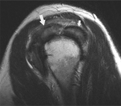
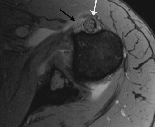
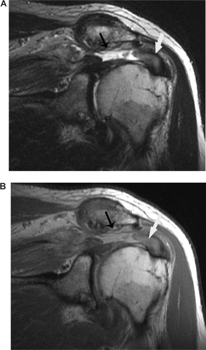
Xanthomatous infiltration may rarely affect the rotator cuff muscles and long head of the biceps tendon. It is the deposition of cholesterol within the rotator cuff muscles and long head of the biceps tendon resulting from hyperlipidemia, specifically high triglyceride and total cholesterol levels. As more commonly seen with xanthomatous infiltration and tear of the Achilles tendon, there may also be an association with rotator cuff tendon deposition and tear. MRI images of xanthomatous infiltration with rotator cuff tear in a 77 year old man with hyperlipidemia are detailed in the following case report.
Keywords: achilles; hyperlipidemia; magnetic resonance imaging; rotator cuff; xanthoma.
Figures
References
-
- Dyslipidemia. Merck & Co Inc; 2008. Merck Manual Professional Edition. Unbound medicine 2008–2009.
-
- Doyle JR. Tendon xanthoma: a physical manifestation of hyperlipidemia. J Hand Surg. 1988 Mar;13(2):238–41. - PubMed
-
- Nakano A, Kinoshita M, Okuda R, Yasuda T, Abe M, Shiomi M. J Orthop Sci. 2006 Jan;11(1):75–80. - PubMed
-
- Sulabha Masih, Tehranzadeh Jamshid. Musculoskeletal Imaging Cases. McGraw Hill; pp. 30–4.pp. 684
-
- Rodriguez C, Goyal M, Wasdahl D. Best Cases from the AFIP. A typical Imaging Features of Bilateral Achilles Tendon Xanthomatosis. Radiographics. 2008;28:2064–8. - PubMed
Publication types
LinkOut - more resources
Full Text Sources
Research Materials
