Patellar skin surface temperature by thermography reflects knee osteoarthritis severity
- PMID: 21151853
- PMCID: PMC2998980
- DOI: 10.4137/CMAMD.S5916
Patellar skin surface temperature by thermography reflects knee osteoarthritis severity
Abstract
Background: Digital infrared thermal imaging is a means of measuring the heat radiated from the skin surface. Our goal was to develop and assess the reproducibility of serial infrared measurements of the knee and to assess the association of knee temperature by region of interest with radiographic severity of knee Osteoarthritis (rOA).
Methods: A total of 30 women (15 Cases with symptomatic knee OA and 15 age-matched Controls without knee pain or knee OA) participated in this study. Infrared imaging was performed with a Meditherm Med2000™ Pro infrared camera. The reproducibility of infrared imaging of the knee was evaluated through determination of intraclass correlation coefficients (ICCs) for temperature measurements from two images performed 6 months apart in Controls whose knee status was not expected to change. The average cutaneous temperature for each of five knee regions of interest was extracted using WinTes software. Knee x-rays were scored for severity of rOA based on the global Kellgren-Lawrence grading scale.
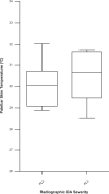
Results: The knee infrared thermal imaging procedure used here demonstrated long-term reproducibility with high ICCs (0.50-0.72 for the various regions of interest) in Controls. Cutaneous temperature of the patella (knee cap) yielded a significant correlation with severity of knee rOA (R = 0.594, P = 0.02).
Conclusion: The skin temperature of the patellar region correlated with x-ray severity of knee OA. This method of infrared knee imaging is reliable and as an objective measure of a sign of inflammation, temperature, indicates an interrelationship of inflammation and structural knee rOA damage.
Keywords: inflammation; infrared imaging; knee; osteoarthritis; thermography.
Figures


References
-
- Braverman IM. The cutaneous microcirculation. J Investig Dermatol Symp Proc. 2000 Dec;5(1):3–9. - PubMed
-
- Bonelli RM, Koltringer P. Autonomic nervous function assessment using thermal reactivity of microcirculation. Clin Neurophysiol. 2000 Oct;111(10):1880–8. - PubMed
-
- Vainer BG. FPA-based infrared thermography as applied to the study of cutaneous perspiration and stimulated vascular response in humans. Phys Med Biol. 2005 Dec 7;50(23):R63–94. - PubMed
-
- Anderson ME, Moore TL, Lunt M, Herrick AL. The ‘distal-dorsal difference’: a thermographic parameter by which to differentiate between primary and secondary Raynaud’s phenomenon. Rheumatology (Oxford) 2007;46(3):533–8. - PubMed
-
- Maeda M, Kachi H, Ichihashi N, Oyama Z, Kitajima Y. The effect of electrical acupuncture-stimulation therapy using thermography and plasma endothelin (ET-1) levels in patients with progressive systemic sclerosis (PSS) J Dermatol Sci. 1998 Jun;17(2):151–5. - PubMed
Grants and funding
LinkOut - more resources
Full Text Sources
Other Literature Sources
Medical
Miscellaneous

