. 2010 Dec 7;5(12):e14246.
doi: 10.1371/journal.pone.0014246.

Intracellular trafficking of guanylate-binding proteins is regulated by heterodimerization in a hierarchical manner

Affiliations

Intracellular trafficking of guanylate-binding proteins is regulated by heterodimerization in a hierarchical manner

Nathalie Britzen-Laurent et al. PLoS One. .

Abstract

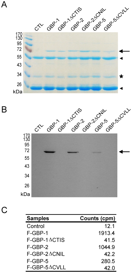
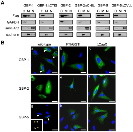
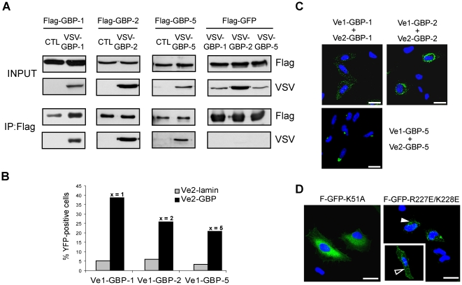
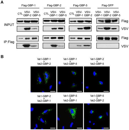
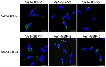
Guanylate-binding proteins (GBPs) belong to the dynamin family of large GTPases and represent the major IFN-γ-induced proteins. Here we systematically investigated the mechanisms regulating the subcellular localization of GBPs. Three GBPs (GBP-1, GBP-2 and GBP-5) carry a C-terminal CaaX-prenylation signal, which is typical for small GTPases of the Ras family, and increases the membrane affinity of proteins. In this study, we demonstrated that GBP-1, GBP-2 and GBP-5 are prenylated in vivo and that prenylation is required for the membrane association of GBP-1, GBP-2 and GBP-5. Using co-immunoprecipitation, yeast-two-hybrid analysis and fluorescence complementation assays, we showed for the first time that GBPs are able to homodimerize in vivo and that the membrane association of GBPs is regulated by dimerization similarly to dynamin. Interestingly, GBPs could also heterodimerize. This resulted in hierarchical positioning effects on the intracellular localization of the proteins. Specifically, GBP-1 recruited GBP-5 and GBP-2 into its own cellular compartment and GBP-5 repositioned GBP-2. In addition, GBP-1, GBP-2 and GBP-5 were able to redirect non-prenylated GBPs to their compartment in a prenylation-dependent manner. Overall, these findings prove in vivo the ability of GBPs to dimerize, indicate that heterodimerization regulates sub-cellular localization of GBPs and underscore putative membrane-associated functions of this family of proteins.


Conflict of interest statement

Competing Interests: The authors have declared that no competing interests exist.

Figures

Figure 1
Figure 1. Human GBP-2 and GBP-5 are prenylated in vivo.
HeLa cells transfected with the empty vector (control) or HeLa expressing Flag-GBP-1, -2 or -5, as well as the respective ΔCaaX mutants, were treated with 200 µCi of [3H]-MVA for 20 h before harvesting. Proteins were extracted, immunoprecipitated with an anti-Flag antibody and separated by 10% SDS-PAGE. (A) Coomassie stained gel. GBPs, GBP mutants are indicated (arrow), as well as the heavy and light chains of the anti-Flag antibody (arrowheads). An unspecific co-precipitated protein is visible (star) (B) Fluorogramm of the gel shown in 1A. The gel was exposed for 3 weeks. (C) Activity measurements of the immunoprecipitates (in cpm, counts per minutes; one fifth of the precipitates was used for quantitative determination).
Figure 2
Figure 2. The CaaX box of GBP-1, GBP-2 and GBP-5 is necessary for the association of the proteins with membranes.
(A) HeLa cells expressing Flag-GBP-1, -2 or -5, as well as the respective ΔCaaX mutants, were fractionated into cytosolic, membranous and nuclear fractions which were analyzed by western blot. GAPDH was used as a marker of the cytosolic fraction, lamin A/C as a marker of the nuclear fraction and cadherin as a marker for the membrane fraction. (B) HeLa cells were transiently transfected with Flag-GFP-GBP-1, Flag-GFP-GBP-2 and Flag-GFP-GBP-5. Cells were either left untreated (a, d, g) or treated with FTI (b, inhibition of GBP-1 farnesylation) and GGTI (e and h, inhibition of GBP-2 and GBP-5 geranylgeranylation, respectively). The presence of GBP-2 in the nucleus is indicated by an asterisk (d and f). Alternatively, HeLa cells were transiently transfected with plasmids expressing GFP-GBP fusion proteins lacking the CaaX box: (c) Flag-GFP-GBP-1ΔCTIS, (f) Flag-GFP-GBP-2ΔCNIL and (i) Flag-GFP-GBP-5ΔCVLL. The insert in (e) shows a co-localization (yellow, arrowhead) between GFP-GBP-5 in green and the Golgi marker GM130 in red. Nuclei were counterstained with DAPI and the Golgi apparatus was stained with an anti-GM130 antibody and an anti-mouse-Alexa546 secondary antibody. Scale bars = 25 µm.
Figure 3
Figure 3. The CaaX motifs of GBPs direct GFP to the Golgi but not to the ER.
(A) HeLa cells were transfected with Flag-GFP-CTIS, -CNIL, –CVLL or a construct containing the polybasic domain and the CaaX box of GBP-1 (GFP-pB-CTIS). Cells were stained with an antibody against the Golgi marker, GM130, and an anti-mouse-AlexaFluor 546 secondary antibody (upper panel) or against the ER marker, calnexin, (lower panel) using an anti-rabbit-AlexaFluor 546 secondary antibody. Nuclei were counterstained with DAPI. Co-localization with the Golgi marker GM130 is indicated by solid arrowheads. Localization at the plasma membrane is indicated by open arrowheads. (B) Prenylation inhibitors abrogate the localization of GFP-CTIS, GFP-CNIL, GFP-CVLL and GFP-pB-CTIS at the Golgi or the plasma membrane. HeLa cells were transiently transfected with Flag-GFP-CTIS, -pB-CTIS, -CNIL and -CVLL. Cells were treated with 10 µM FTI and 10 µM GGTI, as indicated. Scale bars = 25 µm.
Figure 4
Figure 4. The in vivo homodimerization of GBPs differentially influences their intracellular localization.
(A) GBP-1, GBP-2 and GBP-5 are able to homodimerize in vivo. HeLa cells were co-transfected with Flag-GBPs or Flag-GFP together with VSV-GBPs or empty control vector (CTL) as indicated. Protein extracts were immunoprecipitated with an anti-Flag affinity gel and subjected to western blot analysis. For each co-transfection, cell lysates (10 µg, INPUT) and IP eluates (1∶4 for Flag-detection and 3∶4 for VSV detection) were analyzed. (B) Quantitative evaluation of the bi-molecular fluorescence complementation assay. Venus1 plasmids encoding various GBPs were co-transfected with matching Venus2-GBP (black bars) or Venus2-lamin (gray bars) encoding plasmid into HeLa cells. The number of fluorescent cells was determined by FACS analysis after 72 h. (C) Bi-molecular fluorescence complementation assay. HeLa cells were transfected with the plasmids expressing GBP-1, -2 or -5 fused with Venus1 or VSV-Venus2, respectively. Nuclei were counterstained with DAPI. (D) HeLa cells were transfected with different mutants of GBP-1 fused to GFP. Flag-GFP-GBP-1-K51A is defective in nucleotide-dependent oligomerization. Flag-GFP-GBP-1-R227E/K228E is constitutively dimeric and localizes in vesicle-likes structures (solid arrowhead) or at the plasma membrane (open arrowhead). Nuclei were counterstained with DAPI. Scale bars = 25 µm.
Figure 5
Figure 5. The intracellular localization of GBPs is regulated by heterodimerization in a hierarchical manner.
(A) HeLa cells were co-transfected with Flag-GBPs, Flag-GFP and VSV-tagged GBPs or empty control vector (CTL) as indicated. Cell harvesting, Flag-immunoprecipitation and western blot analyses were performed as described in Fig. 3A . (B) HeLa cells were co-transfected with plasmids expressing Venus1-GBP and VSV-Venus2-GBP as indicated. Nuclei were counterstained with DAPI. Scale bars = 25 µm.
Figure 6
Figure 6. Prenylated GBPs recruit non-prenylated GBPs to their subcellular compartments.
HeLa cells were co-transfected with plasmids expressing Venus1-GBP-1, -GBP-2 or -GBP-5 together with plasmids expressing GBP-3 or GBP-4 fused to VSV-Venus2. Nuclei were counterstained with DAPI. Scale bars = 25 µm.

References

    1. Wennerberg K, Rossman KL, Der CJ. The Ras superfamily at a glance. J Cell Sci. 2005;118:843–846. - PubMed
    1. Vetter IR, Wittinghofer A. The guanine nucleotide-binding switch in three dimensions. Science. 2001;294:1299–1304. - PubMed
    1. Vogler O, Barcelo JM, Ribas C, Escriba PV. Membrane interactions of G proteins and other related proteins. Biochim Biophys Acta. 2008;1778:1640–1652. - PubMed
    1. Roskoski R., Jr Protein prenylation: a pivotal posttranslational process. Biochem Biophys Res Commun. 2003;303:1–7. - PubMed
    1. Rocks O, Peyker A, Kahms M, Verveer PJ, Koerner C, et al. An acylation cycle regulates localization and activity of palmitoylated Ras isoforms. Science. 2005;307:1746–1752. - PubMed

Publication types