. 2010 Dec 7;5(12):e14247.
doi: 10.1371/journal.pone.0014247.

IL-6-dependent PGE2 secretion by mesenchymal stem cells inhibits local inflammation in experimental arthritis

Affiliations

IL-6-dependent PGE2 secretion by mesenchymal stem cells inhibits local inflammation in experimental arthritis

Carine Bouffi et al. PLoS One. .

Abstract

Background: Based on their capacity to suppress immune responses, multipotent mesenchymal stromal cells (MSC) are intensively studied for various clinical applications. Although it has been shown in vitro that the immunomodulatory effect of MSCs mainly occurs through the secretion of soluble mediators, the mechanism is still not completely understood. The aim of the present study was to better understand the mechanisms underlying the suppressive effect of MSCs in vivo, using cells isolated from mice deficient in the production of inducible nitric oxide synthase (iNOS) or interleukin (IL)-6 in the murine model of collagen-induced arthritis.

Principal findings: In the present study, we show that primary murine MSCs from various strains of mice or isolated from mice deficient for iNOS or IL-6 exhibit different immunosuppressive potential. The immunomodulatory function of MSCs was mainly attributed to IL-6-dependent secretion of prostaglandin E2 (PGE2) with a minor role for NO. To address the role of these molecules in vivo, we used the collagen-induced arthritis as an experimental model of immune-mediated disorder. MSCs effectively inhibited collagen-induced inflammation during a narrow therapeutic window. In contrast to wild type MSCs, IL-6-deficient MSCs and to a lesser extent iNOS-deficient MSCs were not able to reduce the clinical signs of arthritis. Finally, we show that, independently of NO or IL-6 secretion or Treg cell induction, MSCs modulate the host response by inducing a switch to a Th2 immune response.

Significance: Our data indicate that mscs mediate their immunosuppressive effect via two modes of action: locally, they reduce inflammation through the secretion of anti-proliferative mediators, such as NO and mainly PGE2, and systemically they switch the host response from a Th1/Th17 towards a Th2 immune profile.


Conflict of interest statement

Competing Interests: The authors have declared that no competing interests exist.

Figures

Figure 1
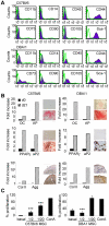
Figure 1. Phenotypic and functional characterization of primary murine MSCs.
A) Immunophenotype of bone marrow-derived MSCs isolated from C57BL/6 and DBA/1 mice. Cells were stained with antibodies against the indicated antigens and analysed by flow cytometry. Representative histograms are shown in black and the respective isotype controls are shown as grey line and are representative of three experiments. B) Differentiation potential of murine MSCs. The differentiation in the 3 lineages is shown by up-regulation of specific markers by RT-qPCR and specific staining. Osteogenesis is characterized by the expression of osteocalcin (OC), alkaline phosphatase (AP) and Alizarin Red S positive staining in differentiation medium (lower panel) versus proliferative medium (upper panel). Adipogenesis is characterized by the expression of peroxysome proliferator-activated receptor (PPAR)-γ; fatty acid binding protein 4 (aP2) and Oil Red O positive staining in differentiation medium (lower panel) versus proliferative medium (upper panel). Chondrogenesis is characterized by the expression of collagen II (Col II) and aggrecan (Agg) and anti-aggrecan positive staining on pellet sections (lower panel) versus unstained control section (upper panel). Results are representative of three independent experiments. C) Immunosuppressive potential of murine C57BL/6 (left panel) and DBA/1 (right panel) MSCs. Allogeneic splenocytes were incubated for three days with ConA without or with various ratios of MSCs. Results are expressed as the percentage of ConA-induced proliferation which was assigned the value of 100%± SEM (n = 3; ***: p<0.005).
Figure 2
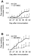
Figure 2. Immunosuppressive effect of syngeneic and allogeneic primary murine MSCs in the CIA model.
A) Evaluation of the severity of arthritis by measurement of the increase of the paw swelling of CIA control mice or mice injected with syngeneic D1 MSCs on day 18 and day 24. Results are representative of two independent experiments. B) Evaluation of the severity of arthritis following injection of allogeneic B6 MSCs at d18 and d24. Results are representative of three independent experiments (*: p<0.05).
Figure 3
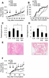
Figure 3. Immunosuppression is dependent of the injection time of primary MSCs in experimental arthritis.
A) Evaluation of the increase of the paw swelling of CIA control mice or mice injected with syngeneic D1 MSCs at various time points. Results are expressed as mean increase ± SEM and are representative of three independent experiments. B) Incidence of arthritis evaluating the percentage of mice with a clinical score >1 (see experimental procedures). C) Radiological score evaluated after X-ray examination on hind paws from mice after euthanasia as assessed in experimental procedures. Results are expressed as the mean ± SEM. D) Histological score evaluated on paraffin sections of hind paws stained by hematoxylin-eosin as assessed in experimental procedures. Results are expressed as the mean ± SEM. E) Representative photomicrographs of joints from CIA control mice (left), or mice injected with MSCs on d18 and d24 (right). Magnification ×5. F) Evaluation of the paw swelling in CIA following injection of D1 MSCs after disease onset. Results are representative of two independent experiments (Statistical analysis showed not significance between groups).
Figure 4
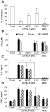
Figure 4. In vitro evaluation of the immunosuppressive potential of MSCs by quantification of soluble mediators.
A) Inhibition of T cell proliferation following co-culture with MSCs. Allogeneic splenocytes (105 cells) were incubated for three days with ConA and when indicated, 5×10⁴ wildtype (wt) C57Bl/6 or MSCs isolated from iNOS−/−, IL-6−/− knock-out C57Bl/6 mice or DBA/1 MSCs were added. Results are expressed as the percentage of ConA-induced proliferation which was assigned the value of 100% ± SEM (n = 3). B) Quantification of NO2 secretion using a modified Griess reagent. C) Quantification of IL-6 secretion by ELISA. D) Quantification of PGE2 secretion by ELISA. Levels of the various soluble mediators were quantified in supernatants of the proliferative assays performed in A) after stimulation with ConA. Parallel assays were performed on cells incubated in the same conditions with the addition of inhibitors specific for NO2 (L-NAME) or PGE2 (indomethacin; Indo). Results are expressed as the mean of 3 independent experiments ± SEM; p values referred to ConA-activated samples (black histograms), if not indicated (*: p<0.05).
Figure 5
Figure 5. Immunosuppressive effect of iNOS−/− and IL-6−/− MSCs in experimental arthritis.
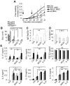
A) Evaluation of the severity of arthritis by measurement of the increase of the paw swelling of control mice (CIA) or mice injected with wt MSCs, iNOS−/− or IL-6−/− MSCs at d18 and d24 (see arrows). Results are expressed as mean increase ± SEM and representative of three independent experiments (*: p<0.05 between wt MSC-treated group and CIA group; #: p≤0.05 between wt MSC-treated group and IL-6−/− MSC-treated group). B) T-cell proliferation assay using splenocytes (105 cells) from the various treated mice incubated with 1, 5 or 25 µg/ml of bCII. C) Determination of the ratio of the bCII-specific IgG1/IgG2a by ELISA in mouse sera. Results are expressed as mean ± SEM. D) Quantification of IL-6 by ELISA in the mouse sera. E) Quantification of cytokines secreted by 2×10⁶ splenocytes cultured in vitro without activation (dashed histograms) or stimulated with 10 µg/ml bCII (black histograms) for 24 h (IFN-γ and IL-17) or 48 h (others) by ELISA. Results are expressed as the mean ± SEM (*: p<0.05; **: p<0.01; ***: p<0.005; NS: not significant).
Figure 6
Figure 6. Induction of a Th2 cellular response after treatment with MSCs.
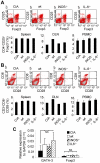
A) Percentage of CD4+CD25+Foxp3+ Treg cells in individual spleen, pooled draining lymph nodes (DLN) and individual peripheral blood mononuclear cells (PBMC) from control CIA or MSC-treated mice at day 31. FACS analyses show CD25+Foxp3+ cells gated on CD4+ T cells in the splenocyte population. B) Percentage of CD3+CD8+CD28 Treg cells in spleen, DLN and PBMC from control CIA or treated mice at day 31. For DLN, cells isolated from all mice in each treated group were pooled resulting in absence of SEM. Results are shown as representative dot plots for 1 mouse per group of mice (upper panel) and histograms in lower panel (% ± SEM), representative of three independent experiments of FACS analyses. C) Relative expression of mRNA level of GATA-3 and Foxp3 transcription factors to GAPDH mRNA in splenocytes using the formula 2−ΔCt. Results are expressed as the mean ± SEM (NS: not significant).

Similar articles

Cited by

References

    1. Vinatier C, Mrugala D, Jorgensen C, Guicheux J, Noel D. Cartilage engineering: a crucial combination of cells, biomaterials and biofactors. Trends Biotechnol. 2009;27:307–314. - PubMed
    1. Djouad F, Bouffi C, Ghannam S, Noel D, Jorgensen C. Mesenchymal stem cells: innovative therapeutic tools for rheumatic diseases. Nat Rev Rheumatol. 2009;5:392–399. - PubMed
    1. Spaggiari GM, Capobianco A, Abdelrazik H, Becchetti F, Mingari MC, et al. Mesenchymal stem cells inhibit natural killer-cell proliferation, cytotoxicity, and cytokine production: role of indoleamine 2,3-dioxygenase and prostaglandin E2. Blood. 2008;111:1327–1333. - PubMed
    1. Sato K, Ozaki K, Oh I, Meguro A, Hatanaka K, et al. Nitric oxide plays a critical role in suppression of T-cell proliferation by mesenchymal stem cells. Blood. 2007;109:228–234. - PubMed
    1. Ren G, Zhang L, Zhao X, Xu G, Zhang Y, et al. Mesenchymal stem cell-mediated immunosuppression occurs via concerted action of chemokines and nitric oxide. Cell Stem Cell. 2008;2:141–150. - PubMed

Publication types