. 2010 Dec 7;4(12):e905.
doi: 10.1371/journal.pntd.0000905.

Fusion between Leishmania amazonensis and Leishmania major parasitophorous vacuoles: live imaging of coinfected macrophages

Affiliations

Fusion between Leishmania amazonensis and Leishmania major parasitophorous vacuoles: live imaging of coinfected macrophages

Fernando Real et al. PLoS Negl Trop Dis. .

Abstract

Protozoan parasites of the genus Leishmania alternate between flagellated, elongated extracellular promastigotes found in insect vectors, and round-shaped amastigotes enclosed in phagolysosome-like Parasitophorous Vacuoles (PVs) of infected mammalian host cells. Leishmania amazonensis amastigotes occupy large PVs which may contain many parasites; in contrast, single amastigotes of Leishmania major lodge in small, tight PVs, which undergo fission as parasites divide. To determine if PVs of these Leishmania species can fuse with each other, mouse macrophages in culture were infected with non-fluorescent L. amazonensis amastigotes and, 48 h later, superinfected with fluorescent L. major amastigotes or promastigotes. Fusion was investigated by time-lapse image acquisition of living cells and inferred from the colocalization of parasites of the two species in the same PVs. Survival, multiplication and differentiation of parasites that did or did not share the same vacuoles were also investigated. Fusion of PVs containing L. amazonensis and L. major amastigotes was not found. However, PVs containing L. major promastigotes did fuse with pre-established L. amazonensis PVs. In these chimeric vacuoles, L. major promastigotes remained motile and multiplied, but did not differentiate into amastigotes. In contrast, in doubly infected cells, within their own, unfused PVs metacyclic-enriched L. major promastigotes, but not log phase promastigotes--which were destroyed--differentiated into proliferating amastigotes. The results indicate that PVs, presumably customized by L. major amastigotes or promastigotes, differ in their ability to fuse with L. amazonensis PVs. Additionally, a species-specific PV was required for L. major destruction or differentiation--a requirement for which mechanisms remain unknown. The observations reported in this paper should be useful in further studies of the interactions between PVs to different species of Leishmania parasites, and of the mechanisms involved in the recognition and fusion of PVs.


Conflict of interest statement

The authors have declared that no competing interests exist.

Figures

Figure 1
Figure 1. L. major amastigote PVs did not fuse with L. amazonensis vacuoles.
(A) Macrophages were previously infected for 48 h with L. amazonensis-WT and then superinfected with L. major-DsRed2 amastigotes for additional 72 hours. Image shows a macrophage loaded with Lysotracker (green) and hosting the two parasite species under phase contrast channel (Ph2), fluorescence channels (Lysotracker and DsRed2), and merged channels, respectively. Asterisk indicates L. amazonensis PV and arrowheads indicate L. major-DsRed2 amastigotes (red). Bars = 10 µm. (B) Live multidimensional imaging of coinfected macrophages. Asterisk indicates L. amazonensis-WT PV stained with Lysotracker (green), surrounded by membrane-bound PVs with weak Lysotracker signal which shelter L. major-DsRed2 amastigotes (red). Multidimensional images were constructed by Imaris blend filter and each image represents a rotation of approximately 45°. Bars = 5 µm. (C) Immunolocalization of LAMP1/LAMP2 proteins in superinfected macrophages. Leishmania major amastigotes were sheltered by tight LAMP1/LAMP2-positive PVs (arrowheads), close to large recipient L. amazonensis PVs, indicated by asterisks. Image was acquired 11 days after L. major-DsRed2 amastigote addition. LAMP immunolabeling in red, 2A3-26 antibody (specific for L. amazonensis amastigotes) immunolabeling in green, DAPI staining in blue. Images are disposed as phase contrast (Ph3), phase contrast with RGB fluorescence channels, phase contrast with RG channels and 3D reconstruction of red channel with Imaris blend filter. Bars = 10 µm.
Figure 2
Figure 2. Fusion of L. amazonensis vacuoles with PVs that shelter L. major promastigotes.
(A) Multidimensional imaging of macrophages infected with L. amazonensis-WT for previous 48 hours and superinfected with L. major-DsRed2 metacyclic-enriched promastigotes. Arrowhead indicates L. major-DsRed2 promastigote sheltered by tight PV, weakly stained with Lysotracker (green), interacting with large, Lysotracker-positive, L. amazonensis-WT PV (asterisk). In the first row, merged images of Lysotracker and DsRed2 signals show transfer of L. major-DsRed2 promastigote to L. amazonensis PV; time after promastigote addition is shown (h:mm). The second row shows DsRed2 signal, evidencing the transfer of promastigote by parasite posterior pole. The third row shows Lysotracker signal, showing changes in PV shape (bold arrow) to accommodate the incoming promastigote. Images were constructed using Imaris blend filter. Bars = 10 µm. (B) Immunolocalization of LAMP1 in superinfected macrophages; Leishmania major–GFP procyclic promastigotes (arrowheads) were sheltered by LAMP1-positive chimeric PV (asterisk). Image was acquired 48 h after L. major-GFP promastigote addition. LAMP1 immunolabeling in red, GFP in green, DAPI staining in blue. Three dimensional images, constructed by Imaris blend filter, are disposed as merged RGB fluorescence channels, merged RB channels and red channel. Bars = 10 µm. (C) Percentage of L. major-GFP promastigotes within chimeric PVs in fixed, superinfected macrophages. Samples were fixed 12 and 72 hours after addition of L. major promastigotes from metacyclic-enriched or log phase parasite batches. Columns are representative of 10 microscopic fields (under 100x objective) in triplicate samples. There is no statistical difference in the percentage of procyclic or metacyclic-enriched promastigotes within chimeric PVs at 12 hours post-superinfection. At 72 hours, a higher percentage of promastigotes from log phase superinfection batch within chimeric PVs was observed, comparing to superinfection with metacyclic-enriched batches (Univariate ANOVA, p<0.05).
Figure 3
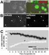
Figure 3. Algorithm-based recognition of L. major-DsRed2 parasites hosted by superinfected macrophages.
(A) Example of an acquired field of macrophages infected for 48 hours with L. amazonensis and superinfected with L. major-DsRed2 metacyclic-enriched promastigotes. First picture is a phase contrast image (Ph2) acquired at 40x magnification, and second is the phase contrast image merged with RG fluorescence channels; Lysotracker in green, L. major-DsRed2 in red. Leishmania amazonensis PVs are indicated by asterisks. (B) Parasite recognition and quantification by Acapella software. The raw data (DsRed2 fluorescence) are shown in the first image and a quantified image is presented in the second (circles represent quantification hits). Examples of parasites recognized by the software are indicated by arrowheads. Bars = 10 µm. (C) Total number of L. major-DsRed2 parasites quantified by software algorithms (black line) and L. major-DsRed2 found within chimeric PVs (white line) quantified by time-lapse videomicrography observation. Acquisition started 2 hours after promastigote addition. Each line represents the mean quantification of 7 microscopic fields (40 x), at logarithmic (base 2) scale, plotted with s.e.m. The total number of promastigotes hosted by superinfected macrophages decreased after approximately 20 hours of experiment. There is a significant increase in the number of L. major parasites found within chimeric PVs in the first 12–20 hours of acquisition (One-way ANOVA with Tamhane's T2, Dunnett's T3 and Games-Howell Post Hoc multiple comparison tests between hourly time points, p<0.05).
Figure 4
Figure 4. L. major promastigotes multiply inside chimeric PVs.
Time-lapse recording of macrophages infected with L. amazonensis-WT for 48 hours and superinfected with metacyclic-enriched L. major-DsRed2 promastigotes. Image acquisition started 6 days after L. major-DsRed2 promastigote addition. Division of L. major-DsRed2 promastigote (arrowheads) inside L. amazonensis-WT PV (asterisk) was documented. The figure shows phase contrast (Ph2) in the first row, DsRed2 signal in the second, and Lysotracker merged with DsRed2 signal in the third. Time after promastigote addition is shown (d:h:min). Scale at 10 µm.
Figure 5
Figure 5. Multidimensional image of metacyclic-enriched L. major-DsRed2 promastigotes in superinfected macrophages.
(A) On the left, multidimensional image of a chimeric PV (asterisk) and L. major-DsRed2 parasites sheltered by unfused donor PVs (arrowheads); Imaris MIP filter. On the right, surface rendering of parasites through DsRed2 channel allowed the software to assign a colorimetric scale to each L. major-DsRed2 parasite: it displays the sphericity parameter, ranging from cyan (less spherical, 0.5) to magenta (more spherical, 0.8); Imaris blend filter. Images were acquired 24 hours after addition of L. major-DsRed2 metacyclic-enriched promastigotes to macrophages. Bar = 10 µm. (B) Sphericity measurements during coinfection, presented by L. major-DsRed2 parasites hosted within unfused donor PVs (magenta lines) or within chimeric PV (blue line). Acquisition of multidimensional images started 12 hours after L. major-DsRed2 metacyclic-enriched promastigotes were added to macrophages.

Similar articles

Cited by

References

    1. Moulder JW. Comparative biology of intracellular parasitism. Microbiol Rev. 1985;49:298–337. - PMC - PubMed
    1. Veras PS, Moulia C, Dauguet C, Tunis CT, Thibon M, et al. Entry and survival of Leishmania amazonensis amastigotes within phagolysosome-like vacuoles that shelter Coxiella burnetii in Chinese hamster ovary cells. Infect Immun. 1995;163:3502–3506. - PMC - PubMed
    1. Rabinovitch M, Veras PS. Cohabitation of Leishmania amazonensis and Coxiella burnetii. Trends Microbiol. 1996;4:158–161. - PubMed
    1. de Chastellier C, Thibon M, Rabinovitch M. Construction of chimeric phagosomes that shelter Mycobacterium avium and Coxiella burnetii (phase II) in doubly infected mouse macrophages: an ultrastructural study. Eur J Cell Biol. 1999;78:580–592. - PubMed
    1. Andreoli WK, Taniwaki NN, Mortara RA. Survival of Trypanosoma cruzi metacyclic trypomastigotes within Coxiella burnetii vacuoles: differentiation and replication within an acidic milieu. Microbes Infect. 2006;8:172–182. - PubMed

Publication types