. 2010 Dec 7;8(12):e1000554.
doi: 10.1371/journal.pbio.1000554.

Domain swapping in allosteric modulation of DNA specificity

Affiliations

Domain swapping in allosteric modulation of DNA specificity

Chad K Park et al. PLoS Biol. .

Abstract

SgrAI is a type IIF restriction endonuclease that cuts an unusually long recognition sequence and exhibits allosteric self-modulation of cleavage activity and sequence specificity. Previous studies have shown that DNA bound dimers of SgrAI oligomerize into an activated form with higher DNA cleavage rates, although previously determined crystal structures of SgrAI bound to DNA show only the DNA bound dimer. A new crystal structure of the type II restriction endonuclease SgrAI bound to DNA and Ca(2+) is now presented, which shows the close association of two DNA bound SgrAI dimers. This tetrameric form is unlike those of the homologous enzymes Cfr10I and NgoMIV and is formed by the swapping of the amino-terminal 24 amino acid residues. Two mutations predicted to destabilize the swapped form of SgrAI, P27W and P27G, have been made and shown to eliminate both the oligomerization of the DNA bound SgrAI dimers as well as the allosteric stimulation of DNA cleavage by SgrAI. A mechanism involving domain swapping is proposed to explain the unusual allosteric properties of SgrAI via association of the domain swapped tetramer of SgrAI bound to DNA into higher order oligomers.


Conflict of interest statement

The authors have declared that no competing interests exist.

Figures

Figure 1
Figure 1. Tetrameric structure of SgrAI.
(A) Dimeric structure of SgrAI bound to primary site DNA (PDB code: 3DVO) . Residues of the swapping domain (1–24) shown in space filling spheres. Residues of the hinge loop (25–30) shown as sticks. Residues 31–339 shown as ribbons, and the bound DNA (black) shown as cartoon. Bound Ca2+ ions shown as black spheres. (B) Tetrameric structure of SgrAI with subunits A, B, G, and H labeled and colored in teal, salmon, slate, and sand, respectively. Each subunit swaps the amino-terminal 24 amino acid residues (shown as space filling spheres) with those of a subunit in an opposing dimer. Residues of the hinge loop (25–30) shown as sticks. Residues 31–339 shown as ribbons, and the bound DNA (black) shown as cartoon. Bound Ca2+ ions shown as black spheres. (C) Ribbon diagram of NgoMIV (PDB code: 1 FIU) (subunits in teal, salmon, slate, and sand) bound to DNA (black) and Mg2+ (black spheres).
Figure 2
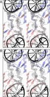
Figure 2. Electron density at swapping hinge loops.
(A) 2Fo-Fc SA omit electron density map at 1 σ for residues 23–31 of subunits B (salmon) and G (slate). Ribbon representation of SgrAI subunits shown in swapped conformation. (B) As in (A) with unswapped conformation.
Figure 3
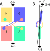
Figure 3. Geometry of SgrAI tetramers.
(A) Positions of 2-fold rotational axes of each dimer in tetramer 1, composed of subunits A, B, G, and H, shown as black lines with a black oval. Subunit colors as in Figure 1A–B. Swapped domains and ordered hinge loops represented by circles and lines with the color of their parent subunit. The hinge loop of subunit A is not ordered. (B) Side view of the tetramer shown in Figure 3A showing the positions of the 2-fold axes. The 2-fold dimeric axis of the upper dimer (subunit G, slate; subunit H, sand; Figure 3A) is 9° from that of the lower dimer (subunit A, teal; subunit B, salmon; Figure 3A) and 10° in tetramer 2.
Figure 4
Figure 4. Swapping Hinge loops.
(A) Close-up of swapped regions of two subunits in the SgrAI tetramer. Residue Arg 31, responsible for recognition of the outer base pair of the primary site recognition sequence, shown as spheres (subunit B, salmon, blue; subunit G, slate, blue). Active site bound Ca2+ ions shown as black spheres and DNA shown in cartoon representation in black. (B) Close-up of interactions at the swapped segments near Pro 27. (C) Same view as in (B) but using unswapped dimer models.
Figure 5
Figure 5. Stimulation of HMWS formation by PCP.
Native PAGE of 1 µM wild type or mutant SgrAI with 1 nM 32P labeled primary or secondary site (18-1 or 18-2), and increasing concentrations of unlabeled precleaved primary site (PCP, 10, 30, 60, 100, 200, 300, 400, 500, 600, 1,000 nM). (A) wild type SgrAI, (B) P27W SgrAI, (C) P27G SgrAI.
Figure 6
Figure 6. Sequences of DNA constructs.
Red, SgrAI primary site recognition sequence; blue, deviation from primary site recognition sequence; arrows, sites of cleavage by SgrAI.

Similar articles

Cited by

References

    1. Bennett M. J, Schlunegger M. P, Eisenberg D. 3D domain swapping: a mechanism for oligomer assembly. Protein Sci. 1995;4:2455–2468. - PMC - PubMed
    1. Liu Y, Eisenberg D. 3D domain swapping: as domains continue to swap. Protein Sci. 2002;11:1285–1299. - PMC - PubMed
    1. Louie G. V, Yang W, Bowman M. E, Choe S. Crystal structure of the complex of diphtheria toxin with an extracellular fragment of its receptor. Mol Cell. 1997;1:67–78. - PubMed
    1. Bennett M. J, Sawaya M. R, Eisenberg D. Deposition diseases and 3D domain swapping. Structure. 2006;14:811–824. - PubMed
    1. Tautz N, Kaluza K, Frey B, Jarsch M, Schmitz G. G, et al. SgrAI, a novel class-II restriction endonuclease from Streptomyces griseus recognizing the octanucleotide sequence 5′-CR/CCGGYG-3′ [corrected]. Nucleic Acids Res. 1990;18:3087. - PMC - PubMed

Publication types

MeSH terms

Associated data