. 2010 Dec 7;8(12):e1000558.
doi: 10.1371/journal.pbio.1000558.

Melanopsin contributions to irradiance coding in the thalamo-cortical visual system

Affiliations

Melanopsin contributions to irradiance coding in the thalamo-cortical visual system

Timothy M Brown et al. PLoS Biol. .

Abstract

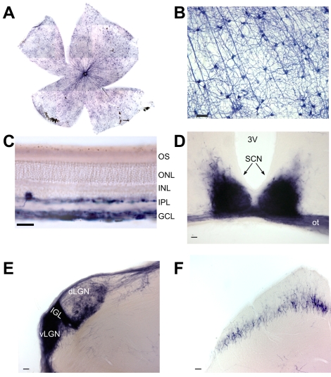
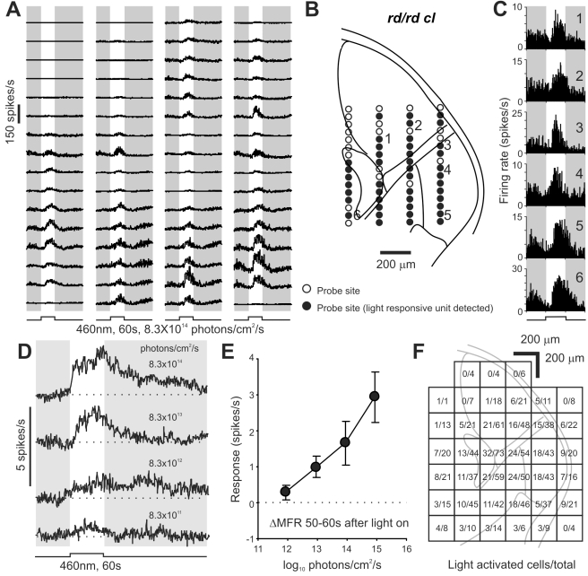
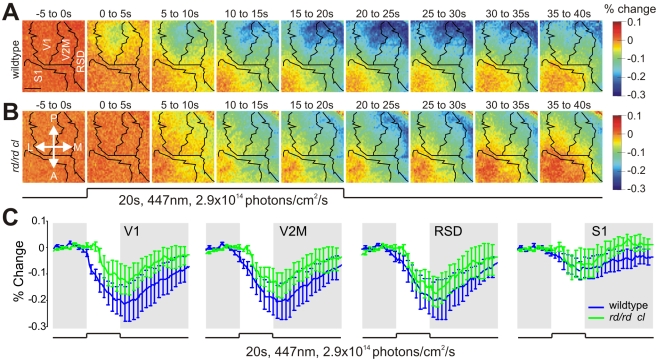
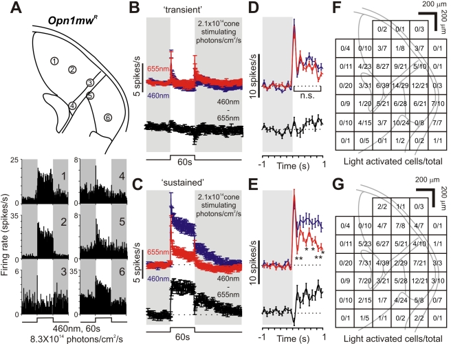
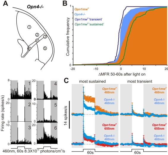
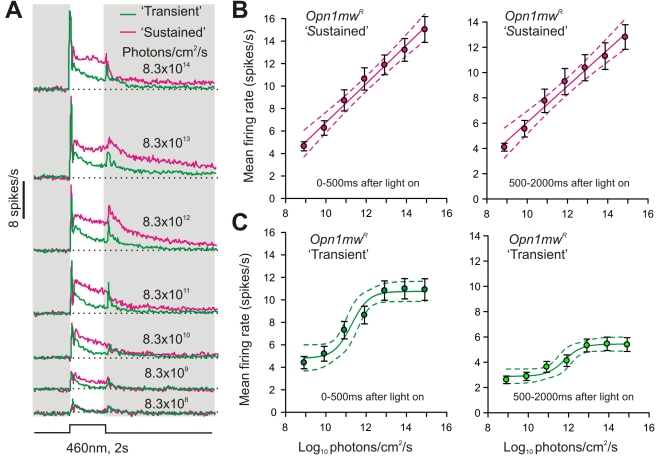
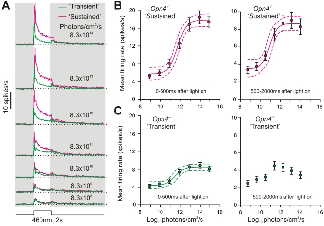
Photoreception in the mammalian retina is not restricted to rods and cones but extends to a subset of retinal ganglion cells expressing the photopigment melanopsin (mRGCs). These mRGCs are known to drive such reflex light responses as circadian photoentrainment and pupillomotor movements. By contrast, until now there has been no direct assessment of their contribution to conventional visual pathways. Here, we address this deficit. Using new reporter lines, we show that mRGC projections are much more extensive than previously thought and extend across the dorsal lateral geniculate nucleus (dLGN), origin of thalamo-cortical projection neurons. We continue to show that this input supports extensive physiological light responses in the dLGN and visual cortex in mice lacking rods+cones (a model of advanced retinal degeneration). Moreover, using chromatic stimuli to isolate melanopsin-derived responses in mice with an intact visual system, we reveal strong melanopsin input to the ∼40% of neurons in the LGN that show sustained activation to a light step. We demonstrate that this melanopsin input supports irradiance-dependent increases in the firing rate of these neurons. The implication that melanopsin is required to accurately encode stimulus irradiance is confirmed using melanopsin knockout mice. Our data establish melanopsin-based photoreception as a significant source of sensory input to the thalamo-cortical visual system, providing unique irradiance information and allowing visual responses to be retained even in the absence of rods+cones. These findings identify mRGCs as a potential origin for aspects of visual perception and indicate that they may support vision in people suffering retinal degeneration.


Conflict of interest statement

The authors have declared that no competing interests exist.

Figures

Figure 1
Figure 1. Genetic labeling of melanopsin RGCs and their projections.
(A) Alkaline phosphatase (AP) stained mRGCs in Opn4Cre;Z/AP mice are uniformly distributed across the retina (1,556±72; mean ± SD, n = 4). (B) AP labels the soma, dendrites, and axons of mRGCs. (C) Labeled cell bodies are restricted to the ganglion cell layer (GCL) and inner nuclear layer (INL), with dendrites in both sublaminae of the inner plexiform layer (IPL). (D–F) AP-stained coronal brain sections demonstrate dense mRGC innervation of the suprachiasmatic nuclei (SCN, D), intergeniculate leaflet (IGL, E), ventral and dorsal lateral geniculate nuclei (v/dLGN, E), and sparse innervation of the superior colliculus (F). 3V, third ventricle; ot, optic tract. Scale bars represent 50 µm (B, D–F) and 25 µm (C).
Figure 2
Figure 2. Melanopsin stimulation activates neurons throughout the lateral geniculate.
(A) Multiunit responses in the LGN of an rd/rd cl mouse showing widespread light activation. Traces are average of four responses and correspond to probe sites shown superimposed on a schematic of the mouse LGN and surrounding thalamus in panel B. Filled circles in (B) indicate probes sites at which a light responsive single unit could be isolated. (C) Representative light responsive single unit firing profiles (numbered sites in B) identified in the dLGN (1,2), IGL/vLGN (3–5), and medial thalamus (6). (D) Average response waveform of light activated cells (n = 344 from 18 mice) to 60 s light pulses of increasing intensity showing kinetics characteristic of the melanopsin photoresponse. (E) Change in mean firing rate (ΔMFR) ± SEM for the data in (D) during the last 10 s of the light pulse. (F) Anatomical distribution of light responsive cells relative to the total number detected in each 200 µm2 grid across all experiments (n = 1,051).
Figure 3
Figure 3. Melanopsin activates the visual cortex.
(A,B) Average pseudo-colored maps of cortical activity in response to 20 s light stimulation in the rd/rd cl and wildtype mice (both n = 4). Green/blue areas on map indicate a decrease in the optical imaging signal, corresponding to neuronal activation. Scale bar represents 1 mm. P, Posterior; M, Medial; A, Anterior; L, Lateral. (C) Time course of the optical imaging signal at different cortical regions (mean ± SEM) in rd/rd cl and wildtype mice. At all but the earliest timepoints the light response rd/rd cl mice accounted for a large proportion of the wildtype response. RSD, retrosplenial dysgranular cortex; S1, somatosensory area 1; V1, visual area 1; V2M, visual area 2 medial.
Figure 4
Figure 4. Sustained responses in the lateral geniculate at high irradiances are not driven by cones.
(A) Responses of six cells recorded from a representative red cone knockin mouse (Opn1mwR; multiunit data in Figure S5) to 60 s blue light pulses. A subset maintained high firing rates throughout light exposure (cell 1,2,6; “sustained”), while others showed only “transient” responses (cell 3–5). Upper panel represents the probe sites of cells 1–6. (B,C) Average (± SEM) response of “transient” (B) and “sustained” (C) cells (n = 134 and 114, respectively, from 10 mice) to 460 nm/655 nm light pulses isoluminant for cones. Black traces represent the subtraction of the 460 nm and 655 nm responses. Responses to 460 nm and 655 nm light were essentially identical in “transient” cells, whereas “sustained” cells consistently exhibited enhanced responses to 460 nm, consistent with a strong melanopsin input. (D,E) Expansion of the initial “On” transient from (B) and (C) at higher temporal resolution (bin size  = 100 ms), responses of “transient” cells (D) were not significantly different while sustained cells (E) showed enhanced short wavelength responses within 100–200 ms following stimulation (paired t tests, n.s. p>0.05, * p<0.05; ** p<0.01). (F,G) Anatomical distribution of “transient” (F) and “sustained” (G) cells relative to the total number of cells detected (based on 542 units). Both cell types were found in approximately equal proportions in both dorsal and ventral portions of the LGN.
Figure 5
Figure 5. Sustained responses are deficient in melanopsin knockout mice.
(A) Responses of six LGN cells from a representative melanopsin knockout mouse (Opn4 /; multiunit data in Figure S6), showing predominantly transient activations. (B) Cumulative frequency distribution of responses to 460 nm stimuli (8.3×10¹⁴ photons/cm2/s) for all light responses in Opn4 / and Opn1mwR (solid areas) and separate “sustained” and “transient” subpopulations (lines) in Opn1mwR, quantified as the change in mean firing rate from baseline during the last 10 s of the light pulse. Opn1mwR was significantly different to Opn4 /, while Opn1mwR “transient” and Opn4 / were not significantly different (Kolmogorov-Smirnov tests; p<0.001 and p>0.05, respectively). (C) Left panel: The response (mean ± SEM) of the 40% of Opn4 / cells with the most sustained light responses was deficient at 460 nm (top) but not 655 nm (bottom) compared to the equivalent population in Opn1mwR mice. Right panel: The remaining 60% of cells in these two genotypes showed equivalent responses (mean ± SEM) at both wavelengths.
Figure 6
Figure 6. Irradiance coding by “sustained” lateral geniculate neurons.
(A) Mean response of “transient” and “sustained” Opn1mwR LGN neurons (n = 134 and 114, respectively) to 2 s blue light pulses. (B,C) Quantification of the firing rate of Opn1mwR “sustained” (B) and “transient” (C) LGN cells during the first 500 ms (left) or remainder (500–2000 ms; right) of the light pulse. Symbols indicate mean (± SEM), and lines indicate mean (±95% CI) of the function that best described the data. Note the linear relationship between firing rate and log irradiance across the full range tested for Opn1mwR “sustained” cells in contrast to the sigmoidal relationship saturating above 1012 photons/cm2/s for Opn1mwR “transient” responses.
Figure 7
Figure 7. Melanopsin is necessary for encoding high irradiances.
(A) Mean response to 2 s blue light pulses from the 40% most “sustained” and remainder 60% most “transient” Opn4 / LGN neurons (as defined by their response to bright 60 s light steps; n = 87 and 130, respectively). (B,C) Quantification of the firing rate of Opn4 / “sustained” (B) or “transient” (C) LGN cells during the first 500 ms (left) or remainder (500–2000 ms; right) of the light pulse. Symbols indicate mean ± SEM, and lines indicate mean ±95% CI of the function that best described the data. Note that, unlike Opn1mwR “sustained” cells, both “sustained” and “transient” Opn4 / populations display sigmoidal relationships between firing rate and irradiance, which saturate above 1012 photons/cm2/s. Data for “transient” Opn4 / cells over 500–2,000 ms of the response were not well fit by either linear or sigmoidal functions.

Comment in

References

    1. Bailes H. J, Lucas R. J. Melanopsin and inner retinal photoreception. Cell Mol Life Sci. 2010;67:99–111. - PMC - PubMed
    1. Belenky M. A, Smeraski C. A, Provencio I, Sollars P. J, Pickard G. E. Melanopsin retinal ganglion cells receive bipolar and amacrine cell synapses. J Comp Neurol. 2003;460:380–393. - PubMed
    1. Perez-Leon J. A, Warren E. J, Allen C. N, Robinson D. W, Lane Brown R. Synaptic inputs to retinal ganglion cells that set the circadian clock. Eur J Neurosci. 2006;24:1117–1123. - PMC - PubMed
    1. Wong K. Y, Dunn F. A, Graham D. M, Berson D. M. Synaptic influences on rat ganglion-cell photoreceptors. J Physiol. 2007;582:279–296. - PMC - PubMed
    1. Viney T. J, Balint K, Hillier D, Siegert S, Boldogkoi Z, et al. Local retinal circuits of melanopsin-containing ganglion cells identified by transsynaptic viral tracing. Curr Biol. 2007;17:981–988. - PubMed

Publication types

LinkOut - more resources