. 2010 Dec 6;5(12):e14232.
doi: 10.1371/journal.pone.0014232.

Atorvastatin improves survival in septic rats: effect on tissue inflammatory pathway and on insulin signaling

Affiliations

Atorvastatin improves survival in septic rats: effect on tissue inflammatory pathway and on insulin signaling

Kelly Lima Calisto et al. PLoS One. .

Erratum in

Retraction in

Abstract

The aim of the present study was to investigate whether the survival-improving effect of atorvastatin in sepsis is accompanied by a reduction in tissue activation of inflammatory pathways and, in parallel, an improvement in tissue insulin signaling in rats. Diffuse sepsis was induced by cecal ligation and puncture surgery (CLP) in male Wistar rats. Serum glucose and inflammatory cytokines levels were assessed 24 h after CLP. The effect of atorvastatin on survival of septic animals was investigated in parallel with insulin signaling and its modulators in liver, muscle and adipose tissue. Atorvastatin improves survival in septic rats and this improvement is accompanied by a marked improvement in insulin sensitivity, characterized by an increase in glucose disappearance rate during the insulin tolerance test. Sepsis induced an increase in the expression/activation of TLR4 and its downstream signaling JNK and IKK/NF-κB activation, and blunted insulin-induced insulin signaling in liver, muscle and adipose tissue; atorvastatin reversed all these alterations in parallel with a decrease in circulating levels of TNF-α and IL-6. In summary, this study demonstrates that atorvastatin treatment increased survival, with a significant effect upon insulin sensitivity, improving insulin signaling in peripheral tissues of rats during peritoneal-induced sepsis. The effect of atorvastatin on the suppression of the TLR-dependent inflammatory pathway may play a central role in regulation of insulin signaling and survival in sepsis insult.


Conflict of interest statement

Competing Interests: The authors have declared that no competing interests exist.

Figures

Figure 1
Figure 1. Effect of atorvastatin on survival in CLP sepsis model.
Male Wistars rats, 8 weeks old, were given saline (Sepsis/Sal, n = 20) or atorvastatin 10 mg/kg (Sepsis/Ator, n = 20), 3 h and once a day after CLP. Survival of the rats was monitored at intervals of 12 h for 15 days. The overall difference in survival rate between the groups with and without atorvastatin was significant (P<0.0001) (A). Fasting blood glucose (B). Fasting insulin levels (C). Glucose disappearance rate (D). HOMA-IR index (E). Serum levels of TNF-α (F) and IL-6 (G). Data are presented as means and S.E. of six to eight rats per group. *P<0.05 (Sepsis saline vs. all others groups).
Figure 2
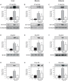
Figure 2. Effects of atorvastatin treatment on insulin signaling in the CLP rat.
Representative blots show insulin-induced tyrosine phosphorylation of Insulin Receptor β (IRβ) in liver (A), muscle (B) and adipose (C) of sham and septic rats. Total protein expression of IRβ (A–C, lower panels). Insulin-induced tyrosine phosphorylation of Insulin Receptor Substrate 1 (IRS1) in liver (D), muscle (E) and adipose tissue (F) of sham and septic rats. Total protein expression of IRS1 (D–F, lower panels). Insulin-induced serine phosphorylation of Akt in liver (G), muscle (H) and adipose (I) of sham and septic rats. Insulin-induced threonine phosphorylation and total protein expression of Akt (G–I, lower panels). In this case, blots were stripped and reprobed with β-actin (A–I, lower panels) to confirm equal loading of proteins. Data are presented as means +/− S.E.M from 6–8 rats per group. *P<0.05 (Sepsis/Sal vs. all others groups). IB, immunoblot; CLT: Sham/Saline; ShT: Sham/Atorvastatin; SAL: saline; ATOR: atorvastatin.
Figure 3
Figure 3. To evaluate the association of TLR4 with MyD88, immunoprecipitations were performed with MyD88 antibody followed by immunoblotting with TLR4 specific antibody.
Representative blots show TLR4 activation (upper panels) and expression (lower panels) in liver (A), muscle (B) and adipose tissue (C) of sham and septic rats. IKKβ phosphorylation in liver (D), muscle (E) and adipose (F) of sham and septic rats. Total protein expression of IKKβ (D–F, lower panels). Phosphorylation of IκBα in liver (G), muscle (H) and adipose (I) of sham and septic rats. Data are presented as means ±S.E.M from 6–8 rats per group. *P<0.05 (Sepsis/Sal vs. all other groups); **P<0.001 (Sepsis/Sal vs. control); #P<0.05 (Sepsis/Sal vs. Sepsis/Ator). IB, immunoblot; CLT: Sham/Saline; ShT: Sham/Atorvastatin; SAL: saline; ATOR: atorvastatin.
Figure 4
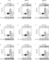
Figure 4. Representative blots show the JNK phosphorylation in liver (A), muscle (B) and adipose tissue (C) of sham and septic rats (upper panels).
Total protein expression of JNK (A–C, lower panels). Phosphorylation of c-jun in liver (D), muscle (E) and adipose tissue (F) of sham and septic rats. Serine 307 Phosphorylation of IRS1 in liver (G), muscle (H) and adipose tissue (I) of sham and septic rats (upper panels). Total protein expression of IRS-1 (G–I, lower panels). Data are presented as means ± S.E.M from 6–8 rats per group. *P<0.05 (Sepsis/Sal vs. all others groups); **P<0.001 (Sepsis/Sal vs. control); #P<0.05 (Sepsis/Sal vs. Sepsis/Ator). IB, immunoblot; CLT: Sham/Saline; ShT: Sham/Atorvastatin; SAL: saline; ATOR: atorvastatin.
Figure 5
Figure 5. Representative blots show the NFkB activation in nuclear fractions of liver (A), muscle (B) and adipose tissue (C) of sham and septic rats.
In this case blots were stripped and reprobed with actin (A–C, lower panels) to confirm equal loading of proteins. Tissue levels of iNOS (D–F) and IL-6 (G–I) expression in liver, muscle and adipose tissue of sham and septic rats. Data are presented as means ± S.E.M from 6–8 rats per group. *P<0.05 (Sepsis/Sal vs. all others groups); **P<0.001 (Sepsis/Sal vs. control); #P<0.05 (Sepsis/Sal vs. Sepsis/Ator). IB, immunoblot; CLT: Sham/Saline; ShT: Sham/Atorvastatin; SAL: saline; ATOR: atorvastatin.
Figure 6
Figure 6. Representative blots show the PERK phosphorylation in liver (A), muscle (B) and adipose tissue (C) of sham and septic rats.
eIF2α phosphorylation (D–F) and ATF6 (G–I) expression in liver, muscle and adipose tissue of sham and septic rats. In this case, blots were stripped and reprobed with actin (A–I, lower panels) to confirm equal loading of proteins. Data are presented as means ± S.E.M from 6–8 rats per group. *P<0.05 (Sepsis/Sal vs. all others groups); #P<0.05 (Sepsis/Sal vs. Sepsis/Ator). IB, immunoblot; CLT: Sham/Saline; ShT: Sham/Atorvastatin; SAL: saline; ATOR: atorvastatin.

Similar articles

Cited by

References

    1. Terblanche M, Almog Y, Rosenson RS, Smith TS, Hackam DG. Statins: panacea for sepsis? Lancet Infect Dis. 2006;6:242–248. - PubMed
    1. Almog Y. Statins, inflammation, and sepsis: hypothesis. Chest. 2003;124:740–743. - PubMed
    1. Maitra SR, Wojnar MM, Lang CH. Alterations in tissue glucose uptake during the hyperglycemic and hypoglycemic phases of sepsis. Shock. 2000;13:379–385. - PubMed
    1. Marik PE, Raghavan M. Stress-hyperglycemia, insulin and immunomodulation in sepsis. Intensive Care Med. 2004;30:748–756. - PubMed
    1. Van den Berghe G. How does blood glucose control with insulin save lives in intensive care? J Clin Invest. 2004;114:1187–1195. - PMC - PubMed

Publication types