. 2010 Dec 7;5(12):e14242.
doi: 10.1371/journal.pone.0014242.

Short-term preservation of porcine oocytes in ambient temperature: novel approaches

Affiliations

Short-term preservation of porcine oocytes in ambient temperature: novel approaches

Cai-Rong Yang et al. PLoS One. .

Abstract

The objective of this study was to evaluate the feasibility of preserving porcine oocytes without freezing. To optimize preservation conditions, porcine cumulus-oocyte complexes (COCs) were preserved in TCM-199, porcine follicular fluid (pFF) and FCS at different temperatures (4°C, 20°C, 25°C, 27.5°C, 30°C and 38.5°C) for 1 day, 2 days or 3 days. After preservation, oocyte morphology, germinal vesicle (GV) rate, actin cytoskeleton organization, cortical granule distribution, mitochondrial translocation and intracellular glutathione level were evaluated. Oocyte maturation was indicated by first polar body emission and spindle morphology after in vitro culture. Strikingly, when COCs were stored at 27.5°C for 3 days in pFF or FCS, more than 60% oocytes were still arrested at the GV stage and more than 50% oocytes matured into MII stages after culture. Almost 80% oocytes showed normal actin organization and cortical granule relocation to the cortex, and approximately 50% oocytes showed diffused mitochondria distribution patterns and normal spindle configurations. While stored in TCM-199, all these criteria decreased significantly. Glutathione (GSH) level in the pFF or FCS group was higher than in the TCM-199 group, but lower than in the non-preserved control group. The preserved oocytes could be fertilized and developed to blastocysts (about 10%) with normal cell number, which is clear evidence for their retaining the developmental potentiality after 3d preservation. Thus, we have developed a simple method for preserving immature pig oocytes at an ambient temperature for several days without evident damage of cytoplasm and keeping oocyte developmental competence.


Conflict of interest statement

Competing Interests: The authors have declared that no competing interests exist.

Figures

Figure 1
Figure 1. Morphologic changes of porcine cumulus-oocyte complexes (COCs) after preservation for 3 d.
a1, a2, b1, b2, c1, c2, When preserved in TCM-199, pFF and FCS at 4°C or 27.5°C, cumulus cells attached tightly to oocytes and displayed no expansion; a3, c3, After preservation in TCM-199 and FCS at 38.5°C, almost all of the cumulus have shed from the oocytes; b3, After pFF preservation at 38.5°C, the whole COCs degenerated and showed black appearance; b2, c2, After preservation in pFF and FCS at 27.5°C, oocytes displayed dark ooplasm with compact cumulus cells. Scale bar  = 200 µm.
Figure 2
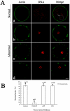
Figure 2. Effect of different media on actin configuration in porcine oocytes after preservation.
A. a, represents the normal actin pattern in porcine oocytes (white arrow pointing at the white circle denotes the germinal vesicle). b, c, and d represent abnormal actin patterns. In b, actin fragments formed an incomplete ring with large punctiform aggregation. In c, small punctiform actin aggregated as an incomplete ring. In d, only some actin aggregated near oocyte membrane. Green, microfilaments; Red, chromatin. Scale bar  = 10 µm. B. Ratios of oocytes with normal actin configuration in control group and different preservation groups. Porcine oocytes were preserved in TCM-199, pFF and FCS at 27.5°C for 3 d, then the actin configuration was stained with FITC-phalloidine. Normal and abnormal actin configuration were detected. The graph shows the mean ± SEM of three independent experiments. The number “n” over the bars means the total treated oocyte in every group. The superscripts a, b over the bars represent values of normal actin configuration that differ significantly (P<0.05).
Figure 3
Figure 3. Effect of different media on CG distribution in porcine oocytes after preservation.
A. a, b, CGs perinuclear area distribution within the porcine oocytes; a, most of the CGs aggregate in the perinuclear area, with a few migrating to the central inner cytoplasm or cortical area; b is intermediate pattern between a and c, with most of the CGs still remaining in perinuclear area and some migrating to the central cytoplasm; c, CGs distributed uniformly in the inner cytoplasmic area, and a few migrated to the cortical area to form a discontinuous ring; d, CGs cortical area distribution; almost all of the CGs have migrated into the cortical area, and a few still remained in the central cytoplasm. Green, cortical granules. Scale bar = 10 µm. (White arrows pointing at the white circle denotes the germinal vesicle). B. Ratios of different CG distribution patterns within the porcine oocytes after preservation in different media. Porcine oocytes were preserved in TCM-199, pFF and FCS at 27.5°C for 3 d, and then CG distribution was stained with FITC-labeled peanut agglutinin. Three categories of CG distribution were detected: perinuclear area; inner cyoplasmic area; cortical area. The graph shows the mean ± SEM of three independent experiments. The number “n” in the bracket means the total treated oocyte in every group. The superscripts a, b over the bars represent values that differ significantly in every categories of CG distribution (P<0.05).
Figure 4
Figure 4. Effects of different media on mitochondria distribution in porcine oocytes after preservation.
A. a, peripheral mitochondrial distribution, with no distribution at the center of the oocytes; b, semi-peripheral mitochondrial distribution; c, mitochondrial relocation and uniform distribution in the inner region of oocyte cytoplasm. Red, Mito Tracker Red staining. Scale bar  = 10 µm. B. Ratios of different mitochondria distribution patterns within the porcine oocytes after preservation with different media. Porcine oocytes were preserved in TCM-199, pFF and FCS at 27.5°C for 3 d, and then mitochondria distribution was stained with MitoTracker Red CMXRos. Three categories of mitochondria distribution were detected: peripheral; semi-peripheral; diffuse. The graph shows the mean ± SEM of three independent experiments. The number “n” in the bracket means the total treated oocyte in every group. The superscripts a, b, c over the bars represent values that differ significantly in every categories of mitochondrial distribution (P<0.05).
Figure 5
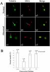
Figure 5. Effects of different preservation media on spindle configuration in porcine oocytes after IVM.
A. a, represents a normal spindle in porcine oocyte. b and c represent abnormal spindle. In b1, c1 arrows indicate abnormal spindle organization distribution. In b2, arrows indicate fragmented and displaced chromosomes. Green, tubulin; Red, chromatin. Scale bar  = 10 µm. B. Normal ratios of spindle configuration in porcine oocytes after preservation in different media following IVM. Porcine oocytes were preserved in TCM-199, pFF and FCS at 27.5°C for 3 d, then spindle was stained with FITC-α-tubulin after culture for 44 h in vitro. Normal and abnormal spindle configuration were detected. The graph shows the mean ± SEM of three independent experiments. The number “n” over the bars means the total treated oocyte in every group. The superscripts a, b over the bars represent values of normal tubulin configuration that differ significantly between groups (P<0.05).

References

    1. Zhou GB, Li N. Cryopreservation of porcine oocytes: recent advances. Mol Hum Reprod. 2009;15:279–285. - PubMed
    1. Didion BA, Pomp D, Martin MJ, Homanics GE, Markert CL. Observations on the cooling and cryopreservation of pig oocytes at the germinal vesicle stage. J Anim Sci. 1990;68:2803–2810. - PubMed
    1. Nagashima H, Kashiwazaki N, Ashman RJ, Grupen CG, Seamark RF, et al. Removal of cytoplasmic lipid enhances the tolerance of porcine embryos to chilling. Biol Reprod. 1994;51:618–622. - PubMed
    1. Liu RH, Sun QY, Li YH, Jiao LH, Wang WH. Maturation of porcine oocytes after cooling at the germinal vesicle stage. Zygote. 2003;11:299–305. - PubMed
    1. Wu C, Rui R, Dai J, Zhang C, Ju S, et al. Effects of cryopreservation on the developmental competence, ultrastructure and cytoskeletal structure of porcine oocytes. Mol Reprod Dev. 2006;73:1454–1462. - PubMed

Publication types