. 2010 Dec 3;5(12):e15466.
doi: 10.1371/journal.pone.0015466.

Cationic amino acid transporters and Salmonella Typhimurium ArgT collectively regulate arginine availability towards intracellular Salmonella growth

Affiliations

Cationic amino acid transporters and Salmonella Typhimurium ArgT collectively regulate arginine availability towards intracellular Salmonella growth

Priyanka Das et al. PLoS One. .

Abstract

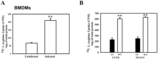
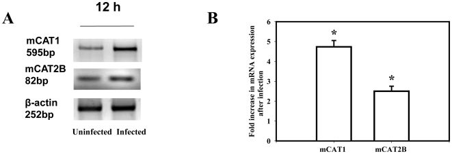
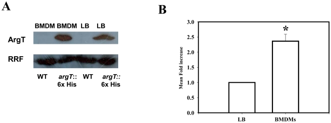
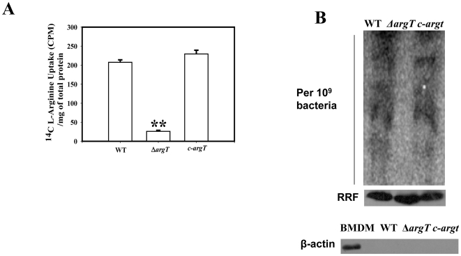
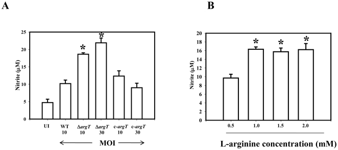
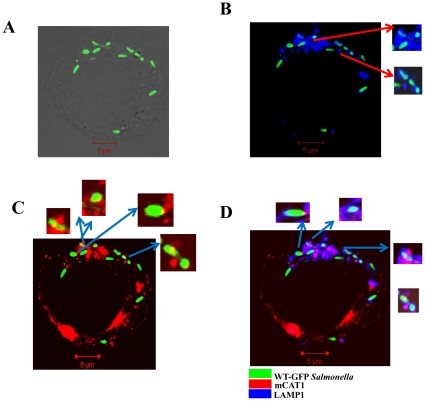
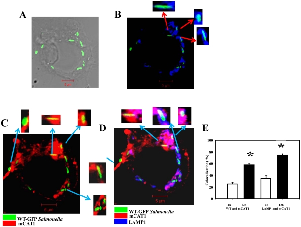
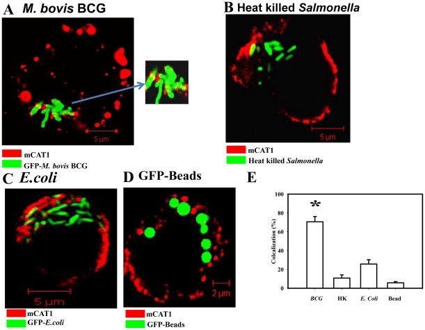
Cationic amino acid transporters (mCAT1 and mCAT2B) regulate the arginine availability in macrophages. How in the infected cell a pathogen can alter the arginine metabolism of the host remains to be understood. We reveal here a novel mechanism by which Salmonella exploit mCAT1 and mCAT2B to acquire host arginine towards its own intracellular growth within antigen presenting cells. We demonstrate that Salmonella infected bone marrow derived macrophages and dendritic cells show enhanced arginine uptake and increased expression of mCAT1 and mCAT2B. We show that the mCAT1 transporter is in close proximity to Salmonella containing vacuole (SCV) specifically by live intracellular Salmonella in order to access the macrophage cytosolic arginine pool. Further, Lysosome associated membrane protein 1, a marker of SCV, also was found to colocalize with mCAT1 in the Salmonella infected cell. The intra vacuolar Salmonella then acquire the host arginine via its own arginine transporter, ArgT for growth. The argT knockout strain was unable to acquire host arginine and was attenuated in growth in both macrophages and in mice model of infection. Together, these data reveal survival strategies by which virulent Salmonella adapt to the harsh conditions prevailing in the infected host cells.


Conflict of interest statement

Competing Interests: The authors have declared that no competing interests exist.

Figures

Figure 1
Figure 1. Salmonella upregulates the arginine transport in Bone Marrow Derived Macrophages (BMDM), in mice liver, spleen and from dendritic cells.
(A) The BMDMs were infected with WT Salmonella at a MOI of 10 and the arginine uptake assay was performed at 12 h post infection, (B) A cohort of 5 mice was inoculated orally with 1×10⁶ CFU of the WT strain and dissected after 5 days post infection. Uninfected (UI) mice were treated with PBS. Liver and Spleen cells from either the infected or UI mice were subjected to the arginine uptake assay. Data represented is one of the three similar experiments. Statistical significance was defined as follows: **, P<0.01 (Student's t test).
Figure 2
Figure 2. Salmonella infection in the Bone Marrow Derived Macrophages (BMDM) increases the expression of the murine cationic amino acid transporter 1 (mCAT1) and murine cationic amino acid transporter 2 (mCAT2B).
(A) The BMDMs were infected with the WT Salmonella at an MOI of 10 and the levels of mCAT1, mCAT2B and β-actin mRNA was examined by Semi quantitative RT-PCR at 12 h post infection. (B) The mean fold increase in mCAT1and mCAT2B transcription after infection from three independent experiments after densitometric analysis and normalization with β-actin is plotted. The levels are compared from the infected to the uninfected macrophages from those experiments. Statistical significance was defined as follows: * P<0.05 (Student's t test).
Figure 3
Figure 3. ArgT is expressed by intracellular Salmonella.
(A) The expression of the His tagged ArgT protein in LB grown and from isolated ArgT:: His knock in bacteria from infected BMDM after running SDS PAGE is shown. The WT control bacteria do not have any His tagged protein in the same size. Ribosome Recycling Factor (RRF) probing was done to equalize the amount of bacterial protein loaded for the SDS PAGE (B) The mean fold increase of the ArgT protein level after infection for 12 h when compared to the LB grown bacteria from three independent experiments after densitometric analysis and normalization with Ribosome Recycling Factor (RRF) was plotted. Statistical significance was defined as follows: * P<0.05, (Student's t test).
Figure 4
Figure 4. Salmonella utilize host derived arginine for protein synthesis.
(A) Arginine uptake assay was performed with 108 numbers of the WT, ΔargT and the argT complemented strain (c- argT) in the late stationery phase of growth. (B) Upper panel, The BMDMs maintained in arginine free DMEM were infected with either the WT, ΔargT or the argT complemented strain (c- argT). After 25 min of infection, radiolabelled arginine was added to the cells. After 12 h of infection equal amounts of the isolated bacterial proteins from 109 bacteria were loaded on SDS page. The gel was exposed in the cassette for 12 h and the incorporated radioactivity was assessed by the phosphor imager scan. Middle panel, Equal loading of the bacteria in all the samples was confirmed by performing anti- Ribosome Recycling Factor (RRF) western blotting. Lower panel, Cellular contamination in the samples were checked by doing β-Actin western blotting. In the positive control with BMDM lysate only the specific band for Actin was observed. Statistical significance was defined as follows: **, P<0.01, **, P<0.01 (Student's t test).
Figure 5
Figure 5. ΔargT Salmonella is attenuated for virulence and shows reduced bacterial proliferation in vivo.
(A) Intracellular survival assay. BMDMs were infected at 10 MOI with the WT, ΔargT or the argT complemented strain (c- argT). Infected macrophages were lysed at 2 and 16 h post infection and the bacterial loads were determined in triplicate. 1×10⁶ bacteria each of the WT or ΔargT or the argT complement strain (c-argT) strains were inoculated orally to a group of 6 male BALB/c mice. After 5th day of infection, homogenized samples of (B) spleen, (C) liver of the infected mice were plated on antibiotic plates and the colonies were counted. Result presented is one of three independent experiments. Statistical significance was defined as follows: *, P<0.05, **, P<0.01 (Mann-Whitney U test).
Figure 6
Figure 6. The ΔargT strain infection in the BMDMs gives rise to high NO response.
(A) BMDMs were infected at 10 MOI with the WT, ΔargT or the argT complemented strain (c- argT) and also with 30 MOI of the ΔargT strain and the argT complemented strain (c- argT) strain. Production of nitrite was determined in the culture supernatants of the infected BMDMs by Griess reaction after 12 h of infection. (B) BMDMs were maintained in 0.5, 1.0, 1.5 or 2.0 mM concentration of L-arginine and infected with the WT Salmonella for 12 h. Production of nitrite was determined in the culture supernatants by Griess reaction. Values are expressed as mean ±SD of one of three independent experiments performed in triplicate. Statistical significance was defined as follows: * P<0.05 (Student's t test).
Figure 7
Figure 7. Intracellular Salmonella starts colocalizing with host mCAT1 in the BMDMs at early time point of infection.
BMDMs were infected with WT Salmonella for 4 h. The sites of colocalizations were detected by immunostaining with an anti-mCAT1 antibody and a Cy5-conjugated secondary antibody and with an anti-LAMP1 antibody and a Cy3-conjugated secondary antibody. Cy3 staining was pseudo-coloured as blue. Samples were analyzed by confocal laser-scanning microscopy and representative images for the localization of the BCG-GFP (green) and the mCAT1 (red) and the LAMP1 (blue) are shown. (A) Representative image for the localization of the WT-GFP (green) in grey scale image. (B) Image for the localization of the WT-GFP (green) and LAMP1 (blue). The colocalization gives rise to cyan colour. Inset: enlarged image of the bacteria containing vacuole. (C) Image for the localization of the CAT1 (red) and WT-GFP (green). The colocalization gives rise to yellow colour. Inset: enlarged image of the bacteria containing vacuole. (D) Image for the localization of the CAT1 (red) and WT-GFP (green) and LAMP1 (blue). LAMP1 and mCAT1 colocalization in the GFP Salmonella gives rise to the same bacteria having both LAMP and mCAT1 immuno-staining, enlarged bacteria are shown in the insets.
Figure 8
Figure 8. Intracellular Salmonella colocalizes with the host mCAT1 in the BMDMs at late time point of infection.
BMDMs were infected with WT Salmonella for 12 h and the staining was done as described earlier. (A) Representative image for the localization of the WT-GFP (green) in grey scale image. (B) Image for the localization of the WT-GFP (green) and LAMP1 (blue). The colocalization gives rise to cyan colour. Inset: enlarged image of the bacteria containing vacuole. (C) Image for the localization of the CAT1 (red) and WT-GFP (green). The colocalization gives rise to yellow colour. Inset: enlarged image of the bacteria containing vacuole. (D) Image for the localization of the CAT1 (red) and WT-GFP (green) and LAMP1 (blue). This gives rise to the same bacteria having both the LAMP and mCAT1 immuno-staining, enlarged bacteria are shown in the insets. The colocalization of the three colours gives rise to white colour. (E) The percent colocalization of WT Salmonella with mCAT1 at 4 (white bar) and 12 h (black bar) post infection. The colocalization (%) of WT-GFP Salmonella with mCAT1 is plotted after counting from 50 fields in three independent experiments. Out of total bacteria the number of yellow bacteria was counted in those fields and colocalization (%) was plotted. The amount of LAMP1 colocalized to the mCAT1 protein is also shown. For this purpose, the colocalization efficiency of Blue that colocalized with Red and gave rise to magenta colour was counted. Statistical significance was tested by comparing the 4 h values with that of the 12 h values an defined as follows:* P<0.05 (Student's t test).
Figure 9
Figure 9. mCAT1 distribution pattern in the BMDMs after infection with various other pathogens or conditions.
(A) Intracellular M. bovis BCG (GFP) colocalizes with the host mCAT1 protein in BMDM after 12 h of infection. The sites of colocalizations were detected by immunostaining with an anti-mCAT1 antibody and a Cy5-conjugated secondary antibody. Samples were analyzed by confocal laser-scanning microscopy and representative images for the localization of the BCG-GFP (green) and the mCAT1 (red) are shown. Inset: enlarged image of the bacteria containing region. (B) Heat killed Salmonella (GFP) after 12 h of infection in BMDM is shown in green and mCAT1 shown in red. Staining was done as described earlier. (C) BMDMS were infected with the dh5a E.coli strain (GFP) at a MOI of 10 for 12 h and staining was done as described earlier. (D) BMDMs were allowed to phagocytose inert latex beads (GFP, Ratio of 1∶25) and fixed after 12 h post phagocytosis. Staining was done as described earlier. (E) The percent colocalization of BCG, Heat killed Salmonella (HK), E.coli or GFP beads (Beads) with mCAT1 are plotted after counting from 50 fields in three independent experiments. Out of total bacteria/bead the total number of yellow bacteria/beads was counted in those fields and colocalization (%) was plotted and the significance was tested after comparing any group with the GFP-Bead's colocalization values. Statistical significance was defined as follows:* P<0.05 (Student's t test).

References

    1. Reeves MW, Evins GM, Heiba AA, Plikaytis BD, Farmer JJ., 3rd Clonal nature of Salmonella typhi and its genetic relatedness to other salmonellae as shown by multilocus enzyme electrophoresis, and proposal of Salmonella bongori comb. nov. J Clin Microbiol. 1989;27:313–320. - PMC - PubMed
    1. Fields PI, Swanson RV, Haidaris CG, Heffron F. Mutants of Salmonella typhimurium that cannot survive within the macrophage are avirulent. Proc Natl Acad Sci U S A. 1986;83:5189–5193. - PMC - PubMed
    1. Chakravortty D, Hansen-Wester I, Hensel M. Salmonella pathogenicity island 2 mediates protection of intracellular Salmonella from reactive nitrogen intermediates. J Exp Med. 2002;195:1155–1166. - PMC - PubMed
    1. Das P, Lahiri A, Lahiri A, Chakravortty D. Modulation of the Arginase Pathway in the Context of Microbial Pathogenesis: A Metabolic Enzyme Moonlighting as an Immune Modulator. PLoS Pathog. 6:e1000899. - PMC - PubMed
    1. Mori M, Gotoh T. Arginine metabolic enzymes, nitric oxide and infection. J Nutr. 2004;134:2820S–2825S; discussion 2853S. - PubMed

Publication types