. 2010 Dec 3;5(12):e14206.
doi: 10.1371/journal.pone.0014206.

TLR4 inhibits mesenchymal stem cell (MSC) STAT3 activation and thereby exerts deleterious effects on MSC-mediated cardioprotection

Affiliations

TLR4 inhibits mesenchymal stem cell (MSC) STAT3 activation and thereby exerts deleterious effects on MSC-mediated cardioprotection

Yue Wang et al. PLoS One. .

Abstract

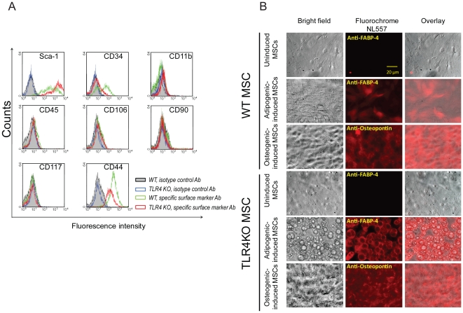
Background: Bone marrow-derived mesenchymal stem cells (MSC) improve myocardial recovery after ischemia/reperfusion (I/R) injury. These effects are mediated in part by the paracrine secretion of angiogenic and tissue growth-promoting factors. Toll-like receptor 4 (TLR4) is expressed by MSC and induces apoptosis and inhibits proliferation in neuronal progenitors as well as many other cell types. It is unknown whether knock-out (KO) of TLR4 will change the paracrine properties of MSC and in turn improve MSC-associated myocardial protection.

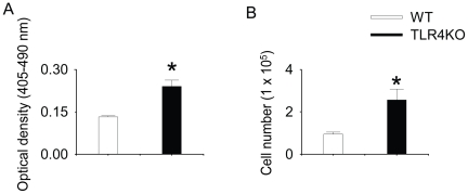
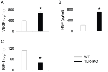
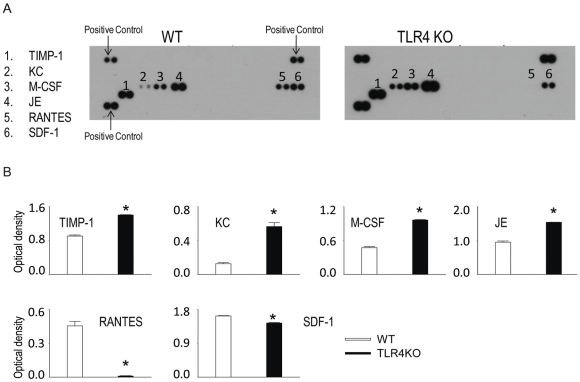
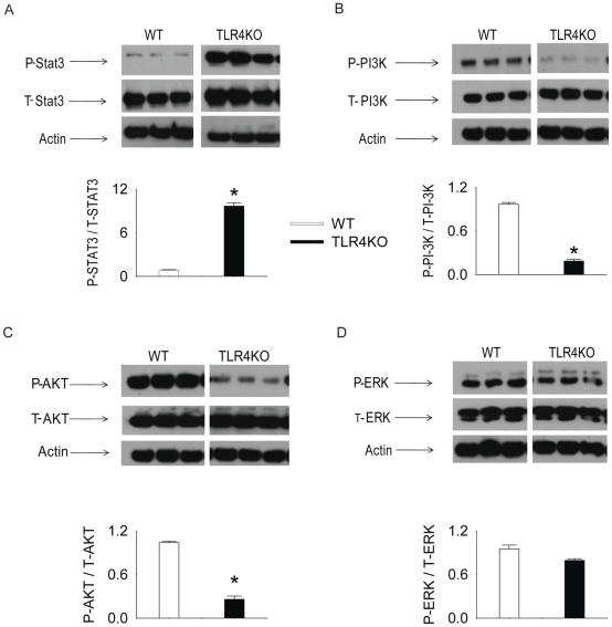
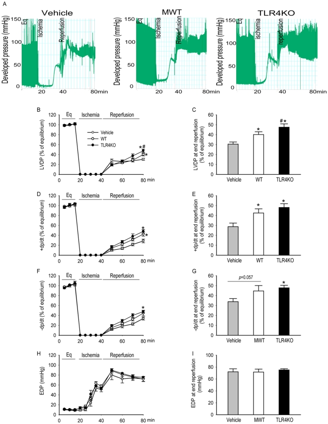
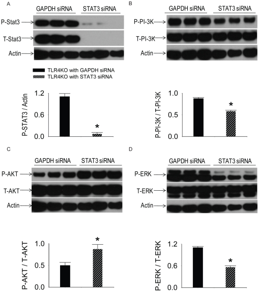
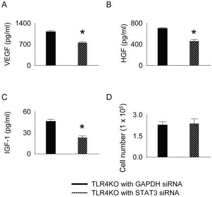
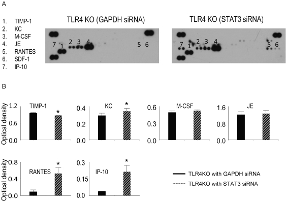
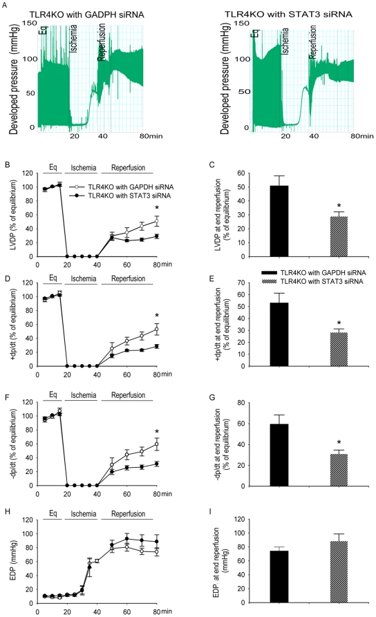
Methodology/principal findings: This study explored the effect of MSC TLR4 on the secretion of angiogenic factors and chemokines in vitro by using ELISA and cytokine array assays and investigated the role of TLR4 on MSC-mediated myocardial recovery after I/R injury in an isolated rat heart model. We observed that MSC isolated from TLR4 KO mice exhibited a greater degree of cardioprotection in a rat model of myocardial I/R injury. This enhanced protection was associated with increased angiogenic factor production, proliferation and differentiation. TLR4-deficiency was also associated with decreased phosphorylation of PI-3K and AKT, but increased activation of STAT3. siRNA targeting of STAT3 resulted in attenuation of the enhanced cardioprotection of TLR4-deficient MSC.

Conclusions/significance: This study indicates that TLR4 exerts deleterious effects on MSC-derived cardioprotection following I/R by a STAT3 inhibitory mechanism.


Conflict of interest statement

Competing Interests: The authors have declared that no competing interests exist.

Figures

Figure 1
Figure 1. MSC express Sca-1 and CD44 and are capable of differentiating into adipocytes and osteocytes.
A: WT and TLR4KO MSC were incubated with specific surface marker antibodies or isotype control antibodies and subjected to flow cytometry analysis. B: MSC in both genotypes were cultured with induction media for 3–4 weeks to induce cell differentiation. The expression of FABP-4 (adipocyte-marker) or osteopontin (osteocyte-marker) was examined using a florescence microscopy (NL557-positive red staining). Representative fields are shown at 100× magnification.
Figure 2
Figure 2. TLR4KO MSC have a higher rate of proliferation than WT MSC.
A: WT and TLR4KO MSC were plated in 96-well plate (5000 cells per well) for 24 h and then incubated with BrdU (10 µM) for an additional 24 h. BrdU incorporation was quantified after incubation. N = 5–6/group. B: WT and TLR4KO MSC were plated in 12-well plates (0.5×10⁵ cell/well). After 24 h, cells were trypsinized and counted using an automatic cell counter. Data are the combination of five independent experiments. Results are mean ± SEM, * p<0.05 vs. WT control.
Figure 3
Figure 3. TLR4KO MSC produced higher levels of VEGF and HGF but lower levels of IGF-1 compared with WT MSC.
WT and TLR4KO MSC were plated in 12-well plates (0.5×10⁵ cell/well). After 24 h, supernatant was collected and the total amounts of VEGF (A), HGF (B) and IGF-1(C) were measured. Data are representative of at least three independent experiments. Results are mean ± SEM; n = 3/group. * p<0.05 vs. WT control.
Figure 4
Figure 4. WT and TLR4KO MSC secrete various chemokines.
WT and TLR4KO MSC were plated in 12-well plates (0.5×10⁵ cell/well). After 24 h, supernatants were collected for cytokine array assay. A: representative cytokine array blots are shown. B: Array signals from images of scanned X-ray films were analyzed and quantified as pixel densities. Data are the combination of two independent experiments. Results are mean ± SEM, n = 4/group, * p<0.05 vs. WT control.
Figure 5
Figure 5. Intracellular kinase activation in WT and TLR4KO MSC.
WT and TLR4KO MSC were plated in 12-well plates (0.05×10⁶ cell/well). After 24 h, cell lysates were collected and the activation of of STAT3 (A), PI-3K (B), AKT (C) and ERK (D) were measured by Western blot. The upper panels in each subfigure show the representative Western blots. The lower panels of each subfigure represent the quantified signal ratio of the activated form to total form (or actin loading control) from the Western blot. The data represent three independent experiments. Results are mean ± SEM, n = 3–4 group, * p<0.05 vs. WT control. Representative blot out of 4 independent experiments are shown.
Figure 6
Figure 6. Intracoronary infusion of TLR4KO MSC provided greater cardioprotection compared to WT MSC.
One million WT or TLR4KO MSC were infused into the coronary circulation prior to global ischemia. The left ventricular function in vehicle (grey, n = 8 rat hearts), WT MSC (white, n = 6), and TLR4KO MSC treated hearts (black, n = 6) is presented. A: representative recording trace from the hearts infused with vehicle, MWT or TLR4KO MSC. The line-graphs represent left ventricular function parameters over time including LVDP (% of equilibrium, B), + dP/dt (% of equilibrium, D), − dP/dt (% of equilibrium, F) and EDP (H). Recovery at end reperfusion is indicated in bar graph form: LVDP (C), + dP/dt (E), −dP/dt (G) and EDP (I). All results are reported as the mean ± SEM. * p<0.05 vs. Vehicle; # p<0.05 vs. WT MSC group.
Figure 7
Figure 7. The role of STAT3 siRNA on the activation of STAT3, PI-3K-AKT and MAPK pathway.
TLR4KO MSC were plated in 12-well plates at 0.5×10⁵cells·well−1·ml−124 h prior to siRNA transfection. The lipofectamine-siRNA complex (100 nM) was added on culture day 2. Medium was then changed after 2 days (day 4 in culture) of transfection. 3 days after transfection (day 5 in culture), cell extracts were prepared for performing Western blots. The activity of STAT3, PI-3K, AKT and ERK was examined by using Western blot. The upper panels in each subfigure show the representative Western blots. The lower panels of each subfigure demonstrate the quantified signals from images of the Western blot. A: STAT3 specific siRNA, but not GAPDH siRNA decreased phosphorylated (P) STAT3 and total (T) STAT3. B–D: STAT3 siRNA suppressed the activation of PI-3K and ERK, but increased the activation of AKT. N = 3 (representative blot out of 6 independent experiments are shown).
Figure 8
Figure 8. TLR4KO MSC ablated with STAT3 produced lower levels of VEGF, HGF and IGF-1 compared with TLR4KO MSC ablated with GAPDH.
TLR4KO MSC were plated in 12-well plates at 0.5×10⁵cells·well−1·ml−124 h prior to siRNA transfection. The lipofectamine-siRNA complex (100 nM) was then added on culture day 2. Medium was then changed after 2 days (day 4 in culture) of transfection. 3 days after transfection (day 5 in culture), the supernatant was collected from TLR4KO MSC either transfected with STAT3 siRNA or GAPDH siRNA and was analyzed by using VEGF, HGF and IGF-1 ELISA. Total amounts of VEGF (A), HGF (B) and IGF-1(C) were examined. siRNA targeting of STAT3 did not change the proliferation of TLR4KO cells (D). Data are representative of at least three independent experiments. Results are mean ± SEM; n = 3/group. * p<0.05 vs. GAPDH ablated group.
Figure 9
Figure 9. siRNA targeting of STAT3 changed the chemokine expression profile in TLR4KO MSC.
TLR4KO MSC were plated in 12-well plates at 0.5×10⁵cells·well−1·ml−124 h prior to siRNA transfection. The lipofectamine-siRNA complex (100 nM) was then added on culture day 2. Medium was then changed after 2 days (day 4 in culture) of transfection. 3 days after transfection (day 5 in culture), the supernatant was collected from TLR4KO MSC either transfected with STAT3 siRNA or GAPDH siRNA and analyzed using a cytokine array assay. A: representative cytokine array blots are shown. B: Array signals from images of scanned X-ray films were analyzed and qualified as pixel densities. Data are the combination of two independent experiments. Results are mean ± SEM, n = 4/group, * p<0.05 vs. GAPDH-ablated TLR4KO group.
Figure 10
Figure 10. siRNA targeting of STAT3 abolished cardioprotection of TLR4KO MSC.
One million TLR4KO MSC transfected with STAT3 siRNA or GAPDH siRNA were infused into the coronary circulation prior to global ischemia. Intracoronary infusion of TLR4KO MSC transfected with GAPDH siRNA provided greater cardioprotection compared to TLR4KO MSC transfected with STAT3 siRNA. The left ventricular function of hearts infused with GAPDH ablated TLR4KO MSC (black, n = 4) and STAT3 ablated TLR4KO MSC (hatched bars, n = 4) is presented. A: representative trace recorded from hearts infused with GAPDH or STAT3 ablated MSC. The line-graphs represent left ventricular function parameters over time including LVDP (% of equilibrium, B), + dP/dt (% of equilibrium, D), − dP/dt (% of equilibrium, F) and EDP (H). Recovery at end reperfusion is indicated in bar graph form: LVDP (C), + dP/dt (E), −dP/dt (G) and EDP (I). All results are reported as the mean ± SEM. * p<0.05 vs. GAPDH group.

References

    1. Haider H, Ashraf M. Bone marrow stem cell transplantation for cardiac repair. Am J Physiol Heart Circ Physiol. 2005;288:H2557–2567. - PubMed
    1. Kudo M, Wang Y, Wani MA, Xu M, Ayub A, et al. Implantation of bone marrow stem cells reduces the infarction and fibrosis in ischemic mouse heart. J Mol Cell Cardiol. 2003;35:1113–1119. - PubMed
    1. Gnecchi M, He H, Liang OD, Melo LG, Morello F, et al. Paracrine action accounts for marked protection of ischemic heart by Akt-modified mesenchymal stem cells. Nat Med. 2005;11:367–368. - PubMed
    1. Togel F, Hu Z, Weiss K, Isaac J, Lange C, et al. Administered mesenchymal stem cells protect against ischemic acute renal failure through differentiation-independent mechanisms. Am J Physiol Renal Physiol. 2005;289:F31–42. - PubMed
    1. Gnecchi M, He H, Noiseux N, Liang OD, Zhang L, et al. Evidence supporting paracrine hypothesis for Akt-modified mesenchymal stem cell-mediated cardiac protection and functional improvement. Faseb J. 2006;20:661–669. - PubMed

Publication types