. 2010 Dec 3;5(12):e14227.
doi: 10.1371/journal.pone.0014227.

Concerted regulation of cGMP and cAMP phosphodiesterases in early cardiac hypertrophy induced by angiotensin II

Affiliations

Concerted regulation of cGMP and cAMP phosphodiesterases in early cardiac hypertrophy induced by angiotensin II

Walid Mokni et al. PLoS One. .

Abstract

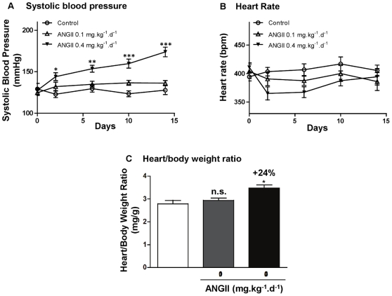
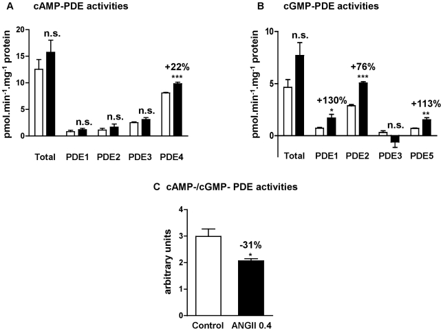
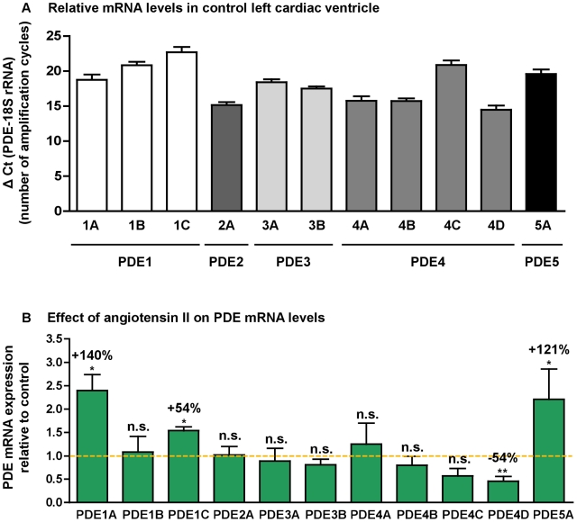
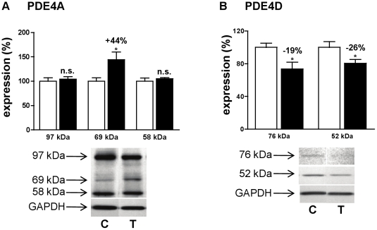
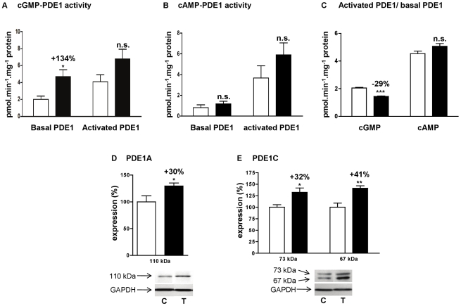
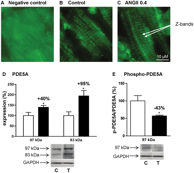
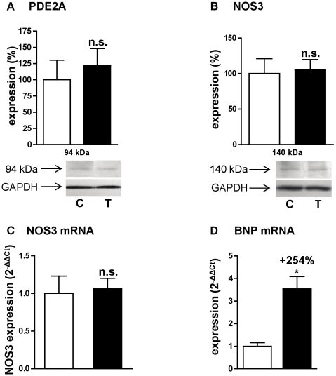
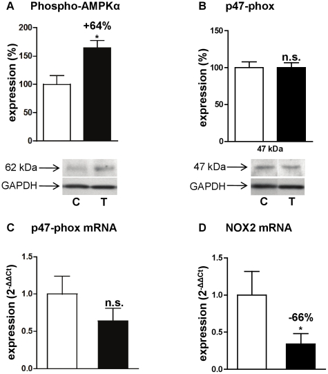
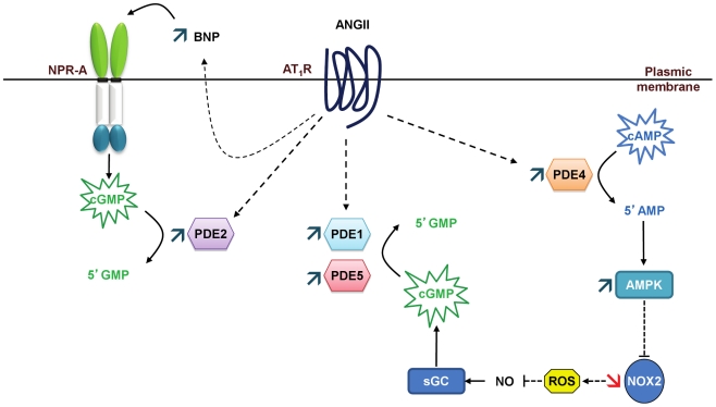
Left ventricular hypertrophy leads to heart failure and represents a high risk leading to premature death. Cyclic nucleotides (cAMP and cGMP) play a major role in heart contractility and cyclic nucleotide phosphodiesterases (PDEs) are involved in different stages of advanced cardiac diseases. We have investigated their contributions in the very initial stages of left ventricular hypertrophy development. Wistar male rats were treated over two weeks by chronic infusion of angiotensin II using osmotic mini-pumps. Left cardiac ventricles were used as total homogenates for analysis. PDE1 to PDE5 specific activities and protein and mRNA expressions were explored.Rats developed arterial hypertension associated with a slight cardiac hypertrophy (+24%). cAMP-PDE4 activity was specifically increased while cGMP-PDE activities were broadly increased (+130% for PDE1; +76% for PDE2; +113% for PDE5) and associated with increased expressions for PDE1A, PDE1C and PDE5A. The cGMP-PDE1 activation by Ca(2+)/CaM was reduced. BNP expression was increased by 3.5-fold, while NOX2 expression was reduced by 66% and AMP kinase activation was increased by 64%. In early cardiac hypertrophy induced by angiotensin II, all specific PDE activities in left cardiac ventricles were increased, favoring an increase in cGMP hydrolysis by PDE1, PDE2 and PDE5. Increased cAMP hydrolysis was related to PDE4. We observed the establishment of two cardioprotective mechanisms and we suggest that these mechanisms could lead to increase intracellular cGMP: i) increased expression of BNP could increase "particulate" cGMP pool; ii) increased activation of AMPK, subsequent to increase in PDE4 activity and 5'AMP generation, could elevate "soluble" cGMP pool by enhancing NO bioavailability through NOX2 down-regulation. More studies are needed to support these assumptions. Nevertheless, our results suggest a potential link between PDE4 and AMPK/NOX2 and they point out that cGMP-PDEs, especially PDE1 and PDE2, may be interesting therapeutic targets in preventing cardiac hypertrophy.


Conflict of interest statement

Competing Interests: The authors have declared that no competing interests exist.

Figures

Figure 1
Figure 1. Effects of angiotensin II treatment on rat physiological parameters.
Systolic blood pressure (A), heart rate (B) and heart/body weight ratio (C). Rats were treated with angiotensin II at 0.1 mg.kg−1.d−1 (ANGII 0.1; n = 8) or 0.4 mg.kg−1.d−1 (ANGII 0.4; n = 13). Effects were expressed in comparison with control rats (n = 6). n.s.: not significant; *: P<0.05; **: P<0.01; ***: P<0.001.
Figure 2
Figure 2. Effects of angiotensin II treatment on left cardiac ventricle PDE specific activities.
cAMP-PDE specific activities and contribution of PDE2, PDE3 and PDE4 (A). cGMP-PDE specific activities and contributions of PDE1, PDE2, PDE3 and PDE5 (B). cAMP-PDE over cGMP-PDE total activity ratio (C). Specific activities were determined on total homogenate and expressed as pmol.min−1.mg−1 of protein. Effects on treated rats (▪; n = 4) were compared with control rats (□; n = 6). n.s.: not significant; *: P<0.05; **: P<0.01; ***: P<0.001.
Figure 3
Figure 3. PDE mRNA distribution in control left cardiac ventricle and effect of angiotensin II treatment.
Relative mRNA levels in control left cardiac ventricle (A). Results were expressed as ΔCt  =  Ct of PDE – Ct of 18S rRNA, which corresponds to the number of amplification cycles needed to detect fluorescence signal, each cycle corresponding to a 2-fold amplification. ΔCt being inversely proportional to initial PDE mRNA level, the fewer is ΔCt, the greater is initial mRNA level. Effects of angiotensin II treatment on PDE mRNA expressions (B). Results were normalized with the 18S rRNA housekeeping gene and expressed as amplification folds relative to control. n.s.: not significant; *: P<0.05; **: P<0.01.
Figure 4
Figure 4. Effect of angiotensin II treatment on cAMP hydrolysis activity.
Effects of angiotensin II treatment at 0.4 mg.kg−1.d−1, on protein expression of PDE4A (A), PDE4D (B). Effects on treated rats (T, ▪; n = 4) were compared with control rats (C, □; n = 4). Results were normalized with GAPDH signal and expressed in percentage of untreated rat. n.s.: not significant; *: P<0.05.
Figure 5
Figure 5. Effect of angiotensin II treatment on PDE1 activity in left cardiac ventricle.
cGMP-PDE1 specific activity (A), cAMP-PDE1 specific activity (B), and PDE1 activated over basal activity ratio (C). Basal PDE1 activity was assessed in presence of EGTA and CaM-activated PDE1 was assessed in presence of Ca2+ and CaM. Effects on treated rats (T, ▪; n = 4) were compared with control rats (C, □□; n = 6). Specific activities were determined on total homogenate and expressed as pmol.min−1.mg−1 of protein. Effects of angiotensin II treatment on protein expression of PDE1A (D) and PDE1C (E). Results were normalized with GAPDH signal and expressed in percentage of untreated rat. n.s.: not significant; *: P<0.05; **: P<0.01; ***: P<0.001.
Figure 6
Figure 6. Effects of angiotensin II treatment on PDE5 expression and intracellular localization.
Immunostaining of PDE5A cellular distribution in left cardiac ventricle: negative control (A), control rats (B) and angiotensin II treated rats (C). PDE5A is localized to Z-bands. Effects of angiotensin II treatment at 0.4 mg.kg−1.d−1 on protein expression of PDE5A (D) and phospho-PDE5A over 97 kDa PDE5A (E). Effects on treated rats (T, ▪; n = 4) were compared with control rats (C, □; n = 4). Results were normalized with GAPDH signal and expressed in percentage of untreated rat. *: P<0.05.
Figure 7
Figure 7. Effects of angiotensin II treatment on PDE2, BNP and NOS.
Expression of PDE2A protein (A), NOS3 protein (B), NOS3 mRNA (C) and BNP mRNA (D). Effects on treated rats (T, ▪; n = 4) were compared with control rats (C, □; n = 4). Western blot results were normalized with GAPDH signal and expressed in percentage of untreated rat. Q-RT-PCR results were normalized with the 18S rRNA housekeeping gene and expressed as amplification folds relative to control. n.s.: not significant; *: P<0.05.
Figure 8
Figure 8. Effects of angiotensin II treatment on AMPK and NADPH oxidase.
Expressions of the phosphorylated α-catalytic subunit of AMPK (A), p47-phox protein (B), p47-phox mRNA (C) and NOX2 mRNA (D). Effects on treated rats (T, ▪; n = 4) were compared with control rats (C, □; n = 4). Western blot results are expressed in percentage after correcting PDE signal with GAPDH signal. Q-RT-PCR results were normalized with the 18S rRNA housekeeping gene and expressed as amplification folds relative to control. n.s.: not significant; *: P<0.05.
Figure 9
Figure 9. Proposed cardioprotective mechanisms that could lead to increase cGMP.
The expression of natriuretic peptides could increase “particulate” cGMP pool, and the activation of AMPK, subsequent to increased PDE4 activity, could increase “soluble” cGMP pool through enhanced NO bioavailability. These increases in cGMP could also have effects on PDE activities: “particulate” cGMP may contribute to further increase PDE2 activation and “soluble” cGMP may further activate PDE5, enhancing even more cGMP hydrolysis.

Similar articles

Cited by

References

    1. Bayes-Genis A, Guindo J, Vinolas X, Tomas L, Elosua R, et al. Cardiac arrhythmias and left ventricular hypertrophy in systemic hypertension and their influences on prognosis. Am J Cardiol. 1995;76:54D–59D. - PubMed
    1. Weber KT, Brilla CG. Structural basis for pathologic left ventricular hypertrophy. Clin Cardiol. 1993;16:II10–14. - PubMed
    1. Serneri GG, Boddi M, Cecioni I, Vanni S, Coppo M, et al. Cardiac angiotensin II formation in the clinical course of heart failure and its relationship with left ventricular function. Circ Res. 2001;88:961–968. - PubMed
    1. Mehta PK, Griendling KK. Angiotensin II cell signaling: physiological and pathological effects in the cardiovascular system. Am J Physiol Cell Physiol. 2007;292:C82–97. - PubMed
    1. Touyz RM, Schiffrin EL. Signal transduction mechanisms mediating the physiological and pathophysiological actions of angiotensin II in vascular smooth muscle cells. Pharmacol Rev. 2000;52:639–672. - PubMed

Publication types