. 2010 Dec 2;6(12):e1001226.
doi: 10.1371/journal.pgen.1001226.

LaeA control of velvet family regulatory proteins for light-dependent development and fungal cell-type specificity

Affiliations

LaeA control of velvet family regulatory proteins for light-dependent development and fungal cell-type specificity

Ozlem Sarikaya Bayram et al. PLoS Genet. .

Abstract

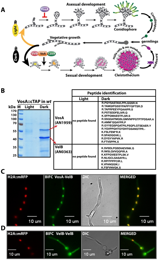
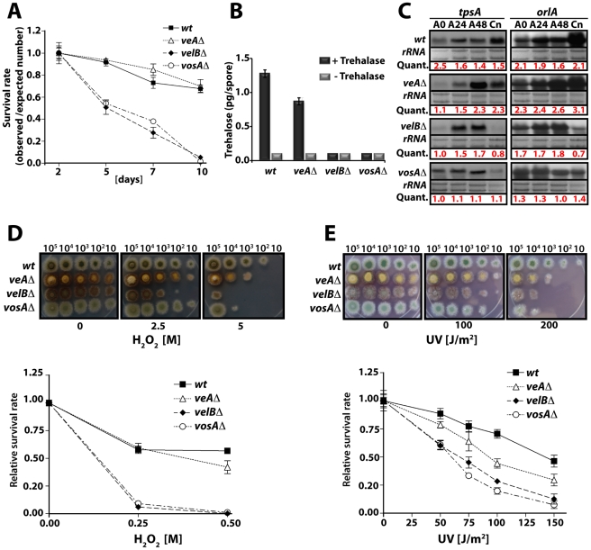
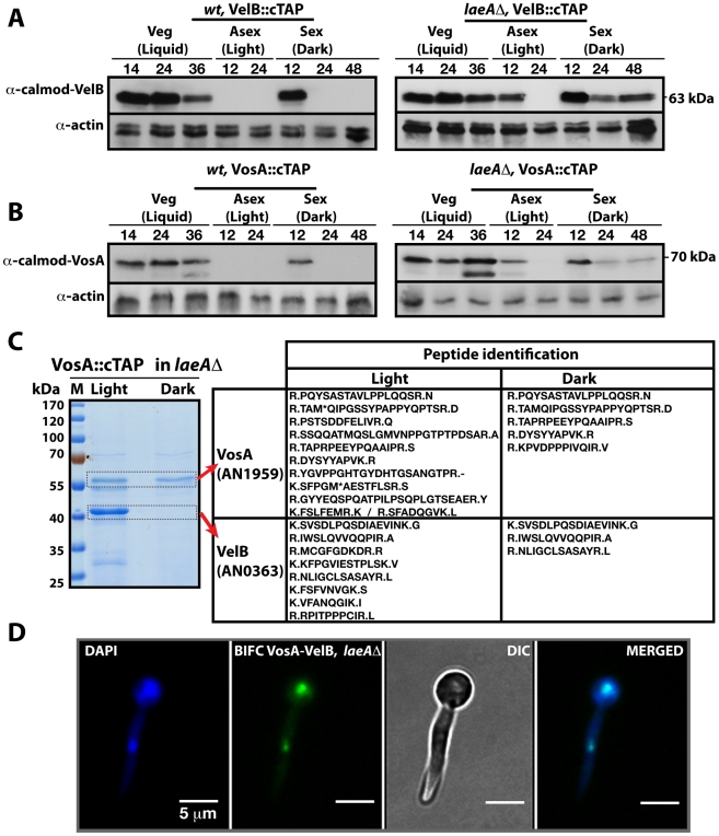
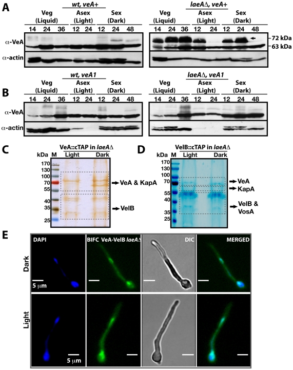
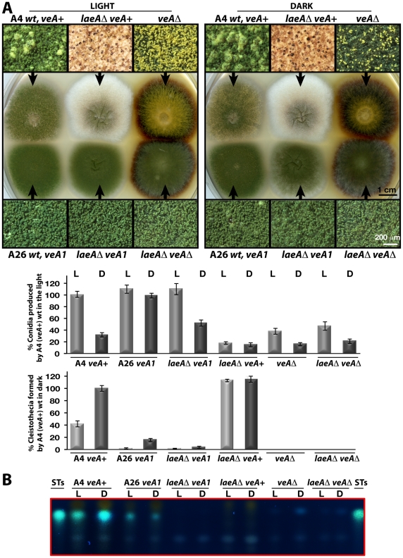
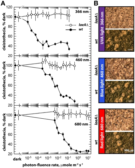
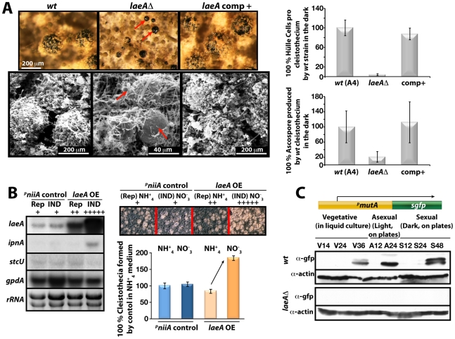
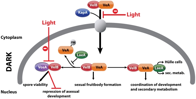
VeA is the founding member of the velvet superfamily of fungal regulatory proteins. This protein is involved in light response and coordinates sexual reproduction and secondary metabolism in Aspergillus nidulans. In the dark, VeA bridges VelB and LaeA to form the VelB-VeA-LaeA (velvet) complex. The VeA-like protein VelB is another developmental regulator, and LaeA has been known as global regulator of secondary metabolism. In this study, we show that VelB forms a second light-regulated developmental complex together with VosA, another member of the velvet family, which represses asexual development. LaeA plays a key role, not only in secondary metabolism, but also in directing formation of the VelB-VosA and VelB-VeA-LaeA complexes. LaeA controls VeA modification and protein levels and possesses additional developmental functions. The laeA null mutant results in constitutive sexual differentiation, indicating that LaeA plays a pivotal role in inhibiting sexual development in response to light. Moreover, the absence of LaeA results in the formation of significantly smaller fruiting bodies. This is due to the lack of a specific globose cell type (Hülle cells), which nurse the young fruiting body during development. This suggests that LaeA controls Hülle cells. In summary, LaeA plays a dynamic role in fungal morphological and chemical development, and it controls expression, interactions, and modification of the velvet regulators.


Conflict of interest statement

The authors have declared that no competing interests exist.

Figures

Figure 1
Figure 1. Life cycle of Aspergillus nidulans and identification of the VosA-associated proteins by tandem affinity purification.
(A) Aspergillus nidulans can grow as a filament (vegetative growth). Light favors asexual development and results in asexual spores (conidiospores) produced by conidiophores. Asexual development is repressed by VosA protein. Darkness favors sexual development and requires the trimeric VelB-VeA-LaeA complex. This leads to fruiting bodies (cleistothecia) nursed by Hülle cells. Meiotically produced sexual spores (ascospores) are formed within the fruiting bodies. White round dots indicate the haploid nuclei of the fungus. (B) SDS-polyacrylamide (10%) gel electrophoresis of TAP enrichment for VosA stained with brilliant blue G. Polypeptides identified from the bands of affinity purification from the light and dark grown cultures are shown (Table S4). (C) Bimolecular fluorescence complementation (BIFC) in vegetative hyphae with enriched nuclear interaction of the VosA-VelB heterodimer. The N-terminal half of the enhanced yellow fluorescent protein (EYFP) fused to the N-terminus of the VosA protein (N-EYFP::VosA) interacts with the C-terminal half of EYFP fused to VelB (C-EYFP::VelB) in vivo. Histone 2A monomeric red fluorescent protein fusion (H2A::mRFP) visualizes the nuclei. (D) BIFC of the VelB-VelB homodimer formation in the cytoplasm and nuclei. N-EYFP::VelB interacts with C-EYFP::VelB.
Figure 2
Figure 2. VelB function in spore viability and trehalose biogenesis.
(A) Viability of wild type and velvet mutant strains conidia grown at 37°C for 2, 5, 7, and 10 days. (B) Amount of trehalose (pg) per conidium in the 2 day old conidia of wild type and the velvet deletion mutants (measured in triplicate). Samples without the trehalase treatment served as controls. (C) Levels of tpsA and orlA transcripts in wild type and velvet mutant strains. Numbers indicate the time (hour) of incubation in post-asexual (A) developmental induction and (Cn) represents conidia. Equal loading of total RNA was evaluated by ethidium bromide staining of rRNA. Quantification of tpsA and orlA expression levels are indicated at the bottom of the blots. Quant: Quantification. (D) Tolerance of the conidia of wild type and velvet mutant strains against H2O2 (see text). (E) Tolerance of the conidia of wild type and velvet mutant strains against ultra violet (UV) irradiation.
Figure 3
Figure 3. LaeA control of VosA and VelB protein levels and the VosA-VelB complex formation.
(A) VelB::cTAP and (B) VosA::cTAP fusion protein levels detected by α-calmodulin antibody during different developmental stages in wild type (wt) and laeAΔ strains at 37°C. α-actin served as internal control. Protein crude extracts (80 µg) were loaded in each lane. (C) Brilliant blue G-stained 10% SDS-polyacrylamid gel of VosA::cTAP and identified polypeptides (Table S5) in laeAΔ strain grown in the light and dark are given. (D) BIFC interaction of the nuclear VosA-VelB complex in laeAΔ strain. N-EYFP::VosA interacts with C-EYFP::VelB. Nuclei were counterstained with DAPI (blue).
Figure 4
Figure 4. VeA-63 kDa and VeA-72 kDa protein levels in wild type and in laeAΔ fungal strains.
(A) The VeA protein levels in wild type (wt) and laeAΔ strains during development (vegetative 14, 24, 36 h in submerged culture, asexual 12, 24 h on plates in the light, sexual 12, 24, and 48 h on plates in the dark at 37°C) by using α-VeA antibodies; α-actin served as internal control. 80 µg total protein was loaded in each lane. (B) The N-terminally truncated VeA1 protein levels in wt and laeAΔ strains. (C) Silver stained 10% SDS- polyacrylamid gel of VeA::cTAP and identified proteins in laeAΔ strain grown in the light and dark (Table S6). (D) SDS-polyacrylamide (10%) gel electrophoresis of VelB::cTAP and associated proteins (in laeAΔ veA+ strain) stained with brilliant blue G (Table S7). (E) BIFC interactions of N-EYFP::VeA and C-EYFP::VelB in laeAΔ fungal cells in light or dark. Nuclei were co-stained by DAPI.
Figure 5
Figure 5. LaeA-VeA as regulators of development and secondary metabolism.
(A) Colony morphologies, quantifications of asexual spore (conidia, in light) and fruiting body (cleistothecia, in dark) formations of (A4) veA+, (A26) veA1, laeAΔ/veA+, laeAΔ/veA1, veAΔ, laeAΔ/veAΔ strains grown on the plates at 37°C for 5 days in the light asexually or in the dark sexually. For the quantification of conidia or cleistothecia, the 5×10 mm2 sectors from 5 independent plates were used and the standard deviations are indicated as vertical bars. veA+ strains conidiation and cleistothecia levels were used as standard (100%). (B) The secondary metabolite sterigmatocystin (ST) production levels of the strains from (A) examined by TLC. 5×10³ conidia were point-inoculated at the center of the plates that were kept either in white light (90 µWm2) or in dark.
Figure 6
Figure 6. Photon fluence-rate response curves for the photoinhibition of cleistothecia formation in wild type and laeAΔ strains.
(A) Petri plates point-inoculated with 5×10³ spores were irradiated with monochromatic light from overhead position at the given photon-fluence rates. wt/veA+; filled circle, laeAΔ/veA+; open circle. Standard errors are represented by vertical lines. (B) Photographs of fruiting bodies (cleistothecia) of wild type (wt) and laeAΔ strains under 366-, 460-, and 680nm light illumination.
Figure 7
Figure 7. LaeA-dependent Hülle cell formation.
(A) Stereo- (top) and scanning electron (SEM) micrographs of wild type (wt), laeAΔ, and laeA complemented strains and quantification of Hülle cells and ascospores per cleistothecium in the dark. Small cleistothecia produced by laeAΔ strain without Hülle cells are indicated by red arrows. Hülle cells and ascospores were counted from 10 different cleistothecia of wt, laeAΔ and laeA complemented strains photographed by SEM. Vertical bars represent standard deviations. Relative values (%) to the numbers of Hülle cells (100–120) or ascospores (2×10⁵) per cleistothecium in wild type are presented. (B) Overproduction of LaeA in veA+ strain increases sexual fruiting body formation in the dark. Growth of wild type (wt) containing an empty niiA promoter plasmid (control), and pniiA::laeA strains. Repressive (5 mM ammonium tartrate) and inducive (10 mM sodium nitrate) conditions were used to confer different levels of the niiA promoter activity. Fruiting body formation of wild type is not affected by these nitrogen sources. The laeA transcript levels were monitored by Northern blot analyses in comparison to ipnA, stcU. gpdA levels and ethidium bromide stained rRNA were used as controls; 20 µg RNA were applied in each lane. Spores (5×10³) were point-inoculated on solid medium and grown at 37°C for 5 days on plates in the dark and cleistothecia were quantified as described . (C) Western blot analysis of Hülle cell specific activity. pmutA::sgfp is specifically expressed in Hülle cells. wt and laeAΔ strains carrying the reporter were grown for indicated time points at 37°C and Western blot with α-gfp, and α-actin as control were performed. 80 µg total protein was applied.
Figure 8
Figure 8. Complexes of velvet family regulatory proteins and LaeA during A. nidulans development.
This model describes the fungal development in dark and the effect of light on nuclear entry and the formation of VosA/VelB complex. VelB primarily enters the nucleus together with VeA and alpha-importin KapA. Then, VelB can be distributed to two distinct complexes. The VosA-VelB dimer can repress asexual spore formation and controls spore maturation and trehalose biogenesis. VeA-VelB can associate with LaeA and the dimeric and/or the trimeric complex controls sexual development. The association of LaeA with the VelB-VeA complex links the secondary metabolism to the development. LaeA controls Hülle cell formation, secondary metabolism and protects VeA against posttranslational modification (PM). VelB is part of the two complexes, VosA-VelB or VelB-VeA.

Similar articles

Cited by

References

    1. Huse JT, Holland EC. Targeting brain cancer: advances in the molecular pathology of malignant glioma and medulloblastoma. Nat Rev Cancer. 2010;10:319–331. - PubMed
    1. Braus GH, Krappmann S, Eckert SE. Sexual Development in Ascomycetes - Fruit Body Formation of Aspergillus nidulans. In: Osiewacz HD, editor. Molecular Biology of Fungal Development. New York, Basel: Marcel Dekker, Inc; 2002. pp. 215–244.
    1. Pöggeler S, Nowrousian M, Kück U. Fruiting-Body Development in Ascomycetes. In: Fischer K, editor. The Mycota I Growth, Differentiation and Sexuality. Heidelberg: Springer-Verlag; 2006. pp. 325–355.
    1. Sohn KT, Yoon KS. Ultrastructural Study on the Cleistothecium Development in Aspergillus nidulans. Mycobiology. 2002;30:117–127.
    1. Keller NP, Turner G, Bennett JW. Fungal secondary metabolism - from biochemistry to genomics. Nat Rev Microbiol. 2005;3:937–947. - PubMed

Publication types

MeSH terms

Substances