Genistein improves neuropathology and corrects behaviour in a mouse model of neurodegenerative metabolic disease
- PMID: 21152017
- PMCID: PMC2995736
- DOI: 10.1371/journal.pone.0014192
Genistein improves neuropathology and corrects behaviour in a mouse model of neurodegenerative metabolic disease
Abstract
Background: Neurodegenerative metabolic disorders such as mucopolysaccharidosis IIIB (MPSIIIB or Sanfilippo disease) accumulate undegraded substrates in the brain and are often unresponsive to enzyme replacement treatments due to the impermeability of the blood brain barrier to enzyme. MPSIIIB is characterised by behavioural difficulties, cognitive and later motor decline, with death in the second decade of life. Most of these neurodegenerative lysosomal storage diseases lack effective treatments. We recently described significant reductions of accumulated heparan sulphate substrate in liver of a mouse model of MPSIIIB using the tyrosine kinase inhibitor genistein.
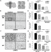
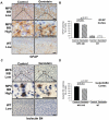
Methodology/principal findings: We report here that high doses of genistein aglycone, given continuously over a 9 month period to MPSIIIB mice, significantly reduce lysosomal storage, heparan sulphate substrate and neuroinflammation in the cerebral cortex and hippocampus, resulting in correction of the behavioural defects observed. Improvements in synaptic vesicle protein expression and secondary storage in the cerebral cortex were also observed.
Conclusions/significance: Genistein may prove useful as a substrate reduction agent to delay clinical onset of MPSIIIB and, due to its multimodal action, may provide a treatment adjunct for several other neurodegenerative metabolic diseases.
Conflict of interest statement
Figures


Similar articles
-
Genistein reduces lysosomal storage in peripheral tissues of mucopolysaccharide IIIB mice.Mol Genet Metab. 2009 Nov;98(3):235-42. doi: 10.1016/j.ymgme.2009.06.013. Epub 2009 Jun 27. Mol Genet Metab. 2009. PMID: 19632871
-
Neuropathology in mouse models of mucopolysaccharidosis type I, IIIA and IIIB.PLoS One. 2012;7(4):e35787. doi: 10.1371/journal.pone.0035787. Epub 2012 Apr 27. PLoS One. 2012. PMID: 22558223 Free PMC article.
-
Early neurodegeneration progresses independently of microglial activation by heparan sulfate in the brain of mucopolysaccharidosis IIIB mice.PLoS One. 2008 May 28;3(5):e2296. doi: 10.1371/journal.pone.0002296. PLoS One. 2008. PMID: 18509511 Free PMC article.
-
Substrate deprivation therapy: a new hope for patients suffering from neuronopathic forms of inherited lysosomal storage diseases.J Appl Genet. 2007;48(4):383-8. doi: 10.1007/BF03195237. J Appl Genet. 2007. PMID: 17998597 Review.
-
[Sanfilippo Syndrome].Vestn Ross Akad Med Nauk. 2015;(4):419-27. Vestn Ross Akad Med Nauk. 2015. PMID: 26710524 Review. Russian.
Cited by
-
Myeloid/Microglial driven autologous hematopoietic stem cell gene therapy corrects a neuronopathic lysosomal disease.Mol Ther. 2013 Oct;21(10):1938-49. doi: 10.1038/mt.2013.141. Epub 2013 Jun 7. Mol Ther. 2013. PMID: 23748415 Free PMC article.
-
The Role of Dimethyl Sulfoxide (DMSO) in Gene Expression Modulation and Glycosaminoglycan Metabolism in Lysosomal Storage Disorders on an Example of Mucopolysaccharidosis.Int J Mol Sci. 2019 Jan 14;20(2):304. doi: 10.3390/ijms20020304. Int J Mol Sci. 2019. PMID: 30646511 Free PMC article.
-
Defects in the medial entorhinal cortex and dentate gyrus in the mouse model of Sanfilippo syndrome type B.PLoS One. 2011;6(11):e27461. doi: 10.1371/journal.pone.0027461. Epub 2011 Nov 9. PLoS One. 2011. PMID: 22096577 Free PMC article.
-
Correction of Huntington's Disease Phenotype by Genistein-Induced Autophagy in the Cellular Model.Neuromolecular Med. 2018 Mar;20(1):112-123. doi: 10.1007/s12017-018-8482-1. Epub 2018 Feb 12. Neuromolecular Med. 2018. PMID: 29435951 Free PMC article.
-
Characterisation of the T cell and dendritic cell repertoire in a murine model of mucopolysaccharidosis I (MPS I).J Inherit Metab Dis. 2013 Mar;36(2):257-62. doi: 10.1007/s10545-012-9508-8. Epub 2012 Jul 7. J Inherit Metab Dis. 2013. PMID: 22773246
References
-
- Poorthuis BJ, Wevers RA, Kleijer WJ, Groener JE, de Jong JG, et al. The frequency of lysosomal storage diseases in The Netherlands. Hum Genet. 1999;105:151–156. - PubMed
-
- Wraith JE. Lysosomal disorders. Semin Neonatol. 2002;7:75–83. - PubMed
-
- Wraith JE. Limitations of enzyme replacement therapy: current and future. J Inherit Metab Dis. 2006;29:442–447. - PubMed
-
- Wynn RF, Wraith JE, Mercer J, O'Meara A, Tylee K, et al. Improved metabolic correction in patients with lysosomal storage disease treated with hematopoietic stem cell transplant compared with enzyme replacement therapy. J Pediatr. 2009;154:609–611. - PubMed
-
- Platt FM, Jeyakumar M. Substrate reduction therapy. Acta. 2008;Paediatr(Suppl 97):88–93. - PubMed
Publication types
MeSH terms
Substances
LinkOut - more resources
Full Text Sources
Other Literature Sources
Medical
Molecular Biology Databases

