. 2010 Dec 1;5(12):e15105.
doi: 10.1371/journal.pone.0015105.

In vitro and in vivo anti-inflammatory activity of 17-O-acetylacuminolide through the inhibition of cytokines, NF-κB translocation and IKKβ activity

Affiliations

In vitro and in vivo anti-inflammatory activity of 17-O-acetylacuminolide through the inhibition of cytokines, NF-κB translocation and IKKβ activity

Mouna Achoui et al. PLoS One. .

Abstract

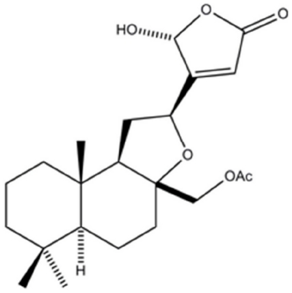
Background and purpose: 17-O-acetylacuminolide (AA), a diterpenoid labdane, was isolated for the first time from the plant species Neouvaria foetida. The anti-inflammatory effects of this compound were studied both in vitro and in vivo.

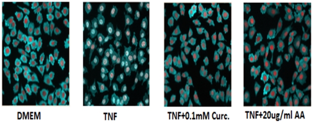
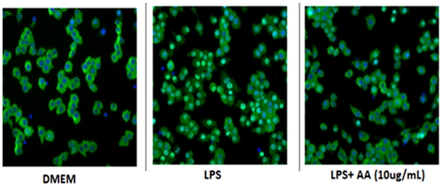
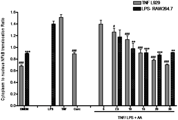
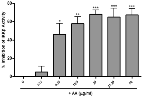
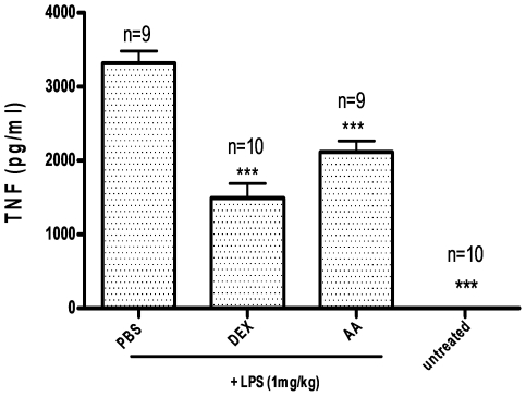
Experimental approach: Plant extracts were initially tested against LPS-stimulated release of tumor necrosis factor alpha (TNF-α) from murine macrophages (RAW264.7 cells). Based on bioassay-guided fractionation, the active compound was identified as AA. AA was tested for its ability to reduce nitric oxide (NO) production, and the inducible nitric oxide synthase (iNOS) expression. The inhibition of a panel of inflammatory cytokines (TNF, IL-1β, IL-6, KC, and GM-CSF) by AA was assessed at the expression and the mRNA levels. Moreover, the effect of AA on the translocation of the transcription factor nuclear factor kappa B (NF-κB) was evaluated in LPS-stimulated RAW264.7 cells and in TNF-stimulated L929 cells. Subsequently, AA was tested in the inhibitor of NF-κB kinase beta (IKKβ) activity assay. Lastly, the anti-inflammatory activity of AA in vivo was evaluated by testing TNF production in LPS-stimulated Balb/c mice.

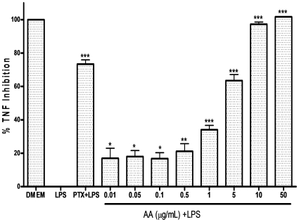
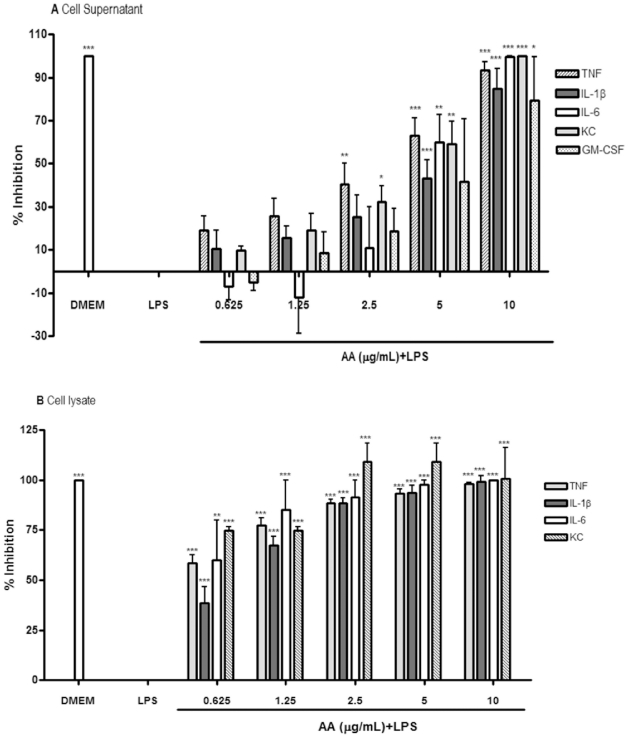
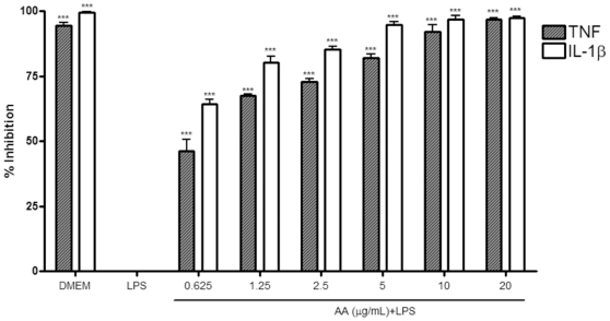
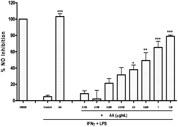
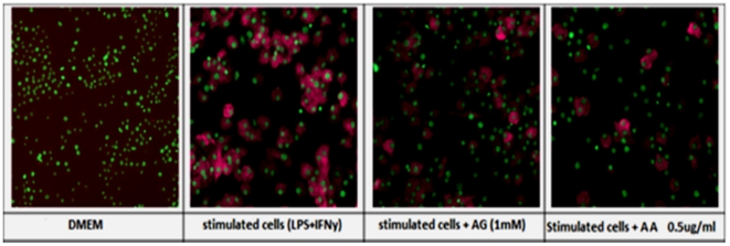
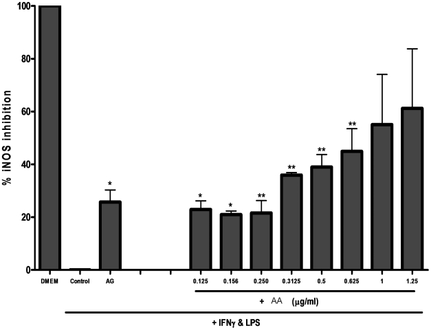
Key results: AA effectively inhibited TNF-α release with an IC(50) of 2.7 µg/mL. Moreover, AA significantly inhibited both NO production and iNOS expression. It significantly and dose-dependently inhibited TNF and IL-1β proteins and mRNA expression; as well as IL-6 and KC proteins. Additionally, AA prevented the translocation of NF-κB in both cell lines; suggesting that it is acting at a post receptor level. This was confirmed by AA's ability to inhibit IKKβ activity, a kinase responsible for activating NF-κB, hence providing an insight on AA's mechanism of action. Finally, AA significantly reduced TNF production in vivo.

Conclusions and implications: This study presents the potential utilization of this compound, as a lead for the development of an anti-inflammatory drug.


Conflict of interest statement

Competing Interests: The authors have declared that no competing interests exist.

Figures

Figure 1
Figure 1. Chemical structure of 17-O-acetylacuminolide [(12S)-17-acetoxy-8a,12-epoxy-16(R)-hydroxylabd-13(14)Z-en-15,16-olide].
Figure 2
Figure 2. The effect of 17-O-acetylacuminolide (AA) on cell viability in RAW264.7 cells.
Cells were pretreated with the indicated doses of AA for 4 hours or were left untreated (DMEM). Data is the average of three independent experiments (±SD), and was analyzed using one way ANOVA with Tukey's post test (** p<0.01, *** p<0.001).
Figure 3
Figure 3. The inhibitory effect of 17-O-acetylacuminolide (AA) on TNF production in RAW264.7 cells.
Cells were pretreated with the indicated doses of AA, or the TNF inhibitor pentoxifylline (PTX). The cells were stimulated with LPS (1 µgmL−1) for four hours, or were left untreated (DMEM).The protein concentration was measured using ELISA. Data is representative of three independent experiments, and was analyzed using one way ANOVA with Tukey's post hoc test (** p<0.01, *** p<0.001).
Figure 4
Figure 4. The inhibitory effect of 17-O-acetylacuminolide (AA) on cytokine release and synthesis in RAW264.7 cells.
Cells were pretreated with the indicated doses of AA. The cells were stimulated with LPS (1 µgmL−1) for 4 hrs, or were left untreated (DMEM). Supernatant (A) and intracellular (B) protein concentrations were measured using Procarta 5-plex cytokine profiling kit from which inhibitions were calculated. Data is representative of three independent experiments, and was analyzed using one-way ANOVA with Tukey's post hoc test (* p<0.05, ** p<0.01, *** p<0.001).
Figure 5
Figure 5. The inhibitory effect of 17-O-acetylacuminolide (AA) on cytokine mRNA.
Cells were treated and mRNA was quantified as described in materials and methods. Data presented is of AA inhibition of TNF and IL-1β mRNA induction and is representative of three independent experiments. Data was analyzed using one-way ANOVA with Tukey's post hoc test (*** p<0.001).
Figure 6
Figure 6. Percentage inhibition of nitric oxide (NO) in RAW264.7.
Cells were pretreated with the indicated doses of 17-O-acetylacuminolide (AA) or with 1 mM of the iNOS inhibitor Aminoguanidine (AG). The cells were stimulated with LPS and IFNγ to activate iNOS synthesis, and eventually NO release. Data presented is the percentage of NO inhibition compared to untreated (DMEM), stimulated cells (control). Data is the average of three independent experiments (±SD), and was analyzed using one way ANOVA with Tukey's post test (* p<0.05, ** p<0.01, *** p<0.001).
Figure 7
Figure 7. Effect of AA on iNOS expression in RAW264.7 cells.
In untreated cells, the nuclei of the cells appear green (hoescht) and iNOS expression was undetected (DMEM). iNOS (DyLight™ 488, magenta), was expressed in the cell cytoplasm of stimulated cells. Pretreatment with aminoguanidine (AG) or 17-O-acetylacuminolide (AA) was able to inhibit iNOS expression in cells stimulated with lipopolysaccharide and interferon gamma (LPS+IFNγ).
Figure 8
Figure 8. Percentage inhibition of inducible nitric oxide synthase (iNOS) in RAW264.7.
Cells were pretreated with the indicated doses of AA or with 1 mM of the iNOS inhibitor Aminoguanidine (AG). The cells were stimulated with LPS and IFNγ to activate iNOS. Data presented is the percentage of iNOS inhibition compared to untreated, stimulated cells (control), and is the average of two experiments. Data was analyzed using one way ANOVA and Tukey's post hoc analysis (* p<0.05, ** p<0.01). Cell count was unaffected at the doses tested (data not shown).
Figure 9
Figure 9. Effect of AA on TNF-stimulated L929 cells.
NF-κB (Dylight™488, light blue); was sequestered in the cytoplasm of cells treated with media only (DMEM), the nuclei of the cells appear red (Hoescht). Upon TNF stimulation (TNF), NF-κB translocated to the nucleus. Pretreatment with curcumin (curc.), or 17-O-acetylacuminolide (AA) was able to prevent NF-κB translocation in the presence of TNF. (AA IC50 = 10.9 µgmL−1).
Figure 10
Figure 10. Effect of AA on LPS-stimulated RAW264.7 cells.
NF-κB (DyLight™ 488, light green), was sequestered in the cytoplasm in cells treated with DMEM alone (untreated), and the nuclei of the cells appear blue (Hoescht). However, NF-κB translocates into the nucleus upon LPS stimulation. Pretreatment with 17-O-acetylacuminolide (AA) was able to prevent NF-κB translocation in the presence of LPS (IC50 = 7.8 µgmL−1).
Figure 11
Figure 11. The inhibitory effect of 17-O-acetylacuminolide (AA) on the increase of nuclear NF-κB intensity.
Cells were pretreated with the indicated doses of AA or with 0.1 mM of the NF-κB inhibitor Curcumin (Curc). L929 cells were stimulated with TNF (1 ngmL−1), whereas RAW264.7 cells were stimulated with LPS (10 ngml−1) to cause NF-κB translocation to the nucleus, or were left untreated (DMEM). Data is the average of three independent experiments (±SD), and was analyzed using one way ANOVA with Tukey's post test. The effect was considered significant when groups were compared to TNF (#) or LPS (*) treated L929 and RAW264.7 cells, respectively. (# p<0.05, ** p<0.01, ###, *** p<0.001).
Figure 12
Figure 12. Effects of 17-O-acetylacuminolide (AA) on IKKβ activity.
Human, recombinant IKKβ (5 ng) was incubated in the presence of increasing concentrations of AA as outlined in the manufacturer's detailed protocol. AA dose-dependently inhibited IKKβ activity with an EC50 of 5.2 µgmL−1. Results are average of three independent experiments ±SD. Data was analyzed using one-way ANOVA, with Tukey's post hoc test (* p<0.05, ** p<0.01, *** p<0.001).
Figure 13
Figure 13. Effect of 17-O-acetylacuminolide (AA) on serum TNF levels in mice.
Mice were pretreated i.p. with either 100 mgkg−1 of AA, 6 mgkg−1 of dexamethasone (DEX) or with Phosphate buffer saline (PBS) and DMSO (untreated) for 30 mins. The mice were then either injected with 1 mgkg−1 lipopolysaccharide (LPS) or with PBS for 90 mins. Blood was withdrawn, and serum TNF was quantified using ELISA. One way ANOVA with Tukey's post analysis was used to calculate the statistical significance among the groups when compared to PBS+LPS group; *** p<0.001.

Similar articles

Cited by

References

    1. Medzhitov R. Origin and physiological roles of inflammation. Nature. 2008;454:428–435. - PubMed
    1. Serhan CN. Resolution Phase of Inflammation: Novel Endogenous Anti-Inflammatory and Proresolving Lipid Mediators and Pathways. Annu Rev Immunol. 2007;25:101–137. - PubMed
    1. Schett G. Rheumatoid arthritis: inflammation and bone loss. Wien Med Wochenschr. 2006;156:34–41. - PubMed
    1. Karin M, Greten FR. NF-kappaB: linking inflammation and immunity to cancer development and progression. Nat Rev Immunol. 2005;5:749–759. - PubMed
    1. Libby P, Ridker P, Maseri A. Inflammation and Atherosclerosis. Circulation. 2002;105:1135–1143. - PubMed

Publication types