. 2010 Nov 18:16:2446-56.

The interaction of unfolding α-lactalbumin and malate dehydrogenase with the molecular chaperone αB-crystallin: a light and X-ray scattering investigation

Affiliations

The interaction of unfolding α-lactalbumin and malate dehydrogenase with the molecular chaperone αB-crystallin: a light and X-ray scattering investigation

Justyn W Regini et al. Mol Vis. .

Abstract

Purpose: The molecular chaperone αB-crystallin is found in high concentrations in the lens and is present in all major body tissues. Its structure and the mechanism by which it protects its target protein from aggregating and precipitating are not known.

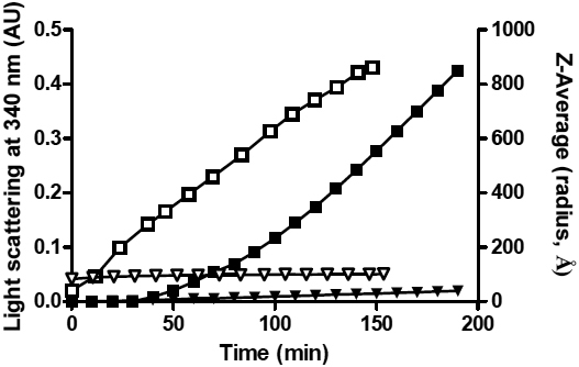
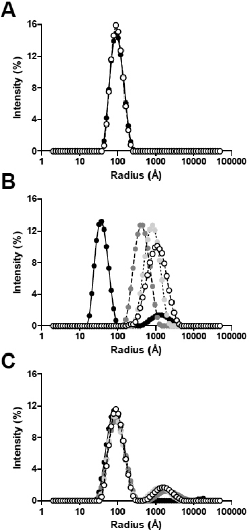
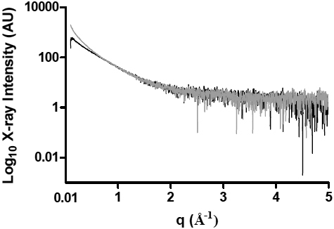
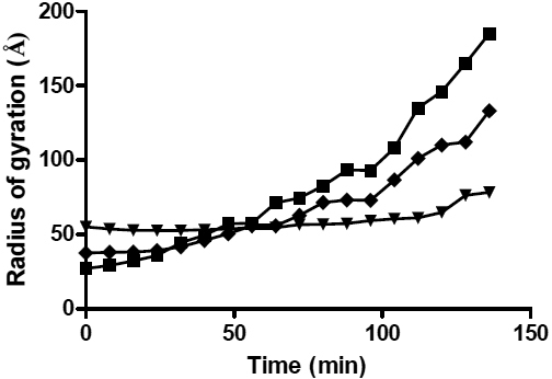
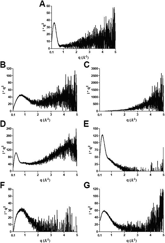
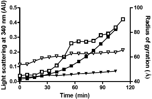
Methods: Dynamic light scattering and X-ray solution scattering techniques were used to investigate structural features of the αB-crystallin oligomer when complexed with target proteins under mild stress conditions, i.e., reduction of α-lactalbumin at 37 °C and malate dehydrogenase when heated at 42 °C. In this investigation, the size, shape and particle distribution of the complexes were determined in real-time following the induction of stress.

Results: Overall, it is observed that the mass distribution, hydrodynamic radius, and spherical shape of the αB-crystallin oligomer do not alter significantly when it complexes with its target protein.

Conclusions: The data are consistent with the target protein being located in the outer protein shell of the αB-crystallin oligomer where it is readily accessible for possible refolding via the action of other molecular chaperones.


Figures

Figure 1
Figure 1
Monitoring the DTT-induced amorphous aggregation of α-LA by light scattering at 340 nm (solid symbols) and dynamic light scattering (open symbols). In both experiments α-LA (2 mg/ml) was incubated at 37 °C in 50 mM phosphate buffer, 100 mM NaCl, 2.5 mM EDTA at pH 7.2 with 20 mM DTT, in the absence (squares) or presence (triangles) of αB-crystallin (1:1 w:w ratio of α-LA: αB-crystallin). The change in light scattering at 340 nm is shown on the left y-axis and the Z-average hydrodynamic radius of particles (Z-average radius, Å) measured by DLS over time is shown on the right y-axis.
Figure 2
Figure 2
Dynamic light scattering measurements of the changes in the distribution of particle sizes (hydrodynamic radii, RH) during the DTT-induced aggregation of α-LA at 37 °C in the absence and presence of αB-crystallin over time. The plots show the relative light scattering intensities (%) of particles of increasing RH (Å) for A αB-crystallin + DTT alone, B DTT-reduced α-LA alone, and C DTT-reduced α-LA in the presence of αB-crystallin (1:1 w:w ratio of α-LA: αB-crystallin). Particle distributions are shown for 0 min (black), 50 min (dark gray), 100 min (light gray) and 150 min (white) following addition of DTT. The RH values used in deriving ρ (see Discussion) are based on the weighted mean of the major peaks in A and C.
Figure 3
Figure 3
The X-ray intensity profiles plotted against the inverse space (q) of α-LA at 37 °C, 32 min (black) and 72 min (gray) after the addition of DTT.
Figure 4
Figure 4
The variation in the radius of gyration (Rg) with time after the addition of 20 mM DTT to α-LA (squares), and in the presence of αB-crystallin at a 1:1 (triangles) and 1:10 (diamonds) w:w ratios to α-LA. The standard deviations associated with these data, which represent the standard deviation from the line of best fit in the Guinier region, are too small to be distinguished in this plot. The Rg values used in deriving ρ (see Discussion) are taken from the final Rg values in this plot.
Figure 5
Figure 5
X-ray intensity profiles plotted against I*q2. Kraty plots are shown for A αB-crystallin alone, α-LA + DTT at B 0 min, and C 136 min, αB-crystallin plus α-LA at a 1:1 w:w ratio at D 0 min and E 136 min, and αB-crystallin plus α-LA at a 1:10 w:w ratio at F 0 min and G 136 min.
Figure 6
Figure 6
Monitoring the thermally-induced amorphous aggregation of MDH by light scattering at 360 nm (solid symbols) and SAXS (open symbols). In both experiments a 2 mg/ml solution of MDH was incubated at 42 °C in 50 mM phosphate buffer, 100 mM NaCl, 2.5 mM EDTA at pH 7.5 in the absence (squares) or presence (triangles) of αB-crystallin (2.5:1.0 w:w ratio of MDH and αB-crystallin for the light scattering experiments and a 1.0:1.0 w:w ratio of MDH and αB-crystallin for the SAXS experiments). The change in light scattering at 360 nm is shown on the left y-axis and the radius of gyration of the samples over time is shown on the right y-axis. The standard deviations associated with the SAXS data, which represent the standard deviation from the line of best fit in the Guinier region, are too small to be distinguished in this plot.

Similar articles

Cited by

References

    1. van Montfort RLM, Basha E, Friedrich KL, Slingsby C, Vierling E. Crystal structure and assembly of a eukaryotic small heat shock protein. Nat Struct Biol. 2001;8:1025–30. - PubMed
    1. Treweek TM, Morris AM, Carver JA. Intracellular protein unfolding and aggregation: The role of small heat-shock chaperone proteins. Aust J Chem. 2003;56:357–67.
    1. Horwitz J. Alpha-crystallin. Exp Eye Res. 2003;76:145–53. - PubMed
    1. Derham BK, Harding JJ. Alpha-crystallin as a molecular chaperone. Prog Retin Eye Res. 1999;18:463–509. - PubMed
    1. Carver JA, Rekas A, Thorn DC, Wilson MR. Small heat-shock proteins and clusterin: intra- and extracellular molecular chaperones with a common mechanism of action and function? IUBMB Life. 2003;55:661–8. - PubMed

Publication types

LinkOut - more resources