. 2010 Nov 30;5(11):e14157.
doi: 10.1371/journal.pone.0014157.

IGF-IR internalizes with Caveolin-1 and PTRF/Cavin in HaCat cells

Affiliations

IGF-IR internalizes with Caveolin-1 and PTRF/Cavin in HaCat cells

Barbara Salani et al. PLoS One. .

Abstract

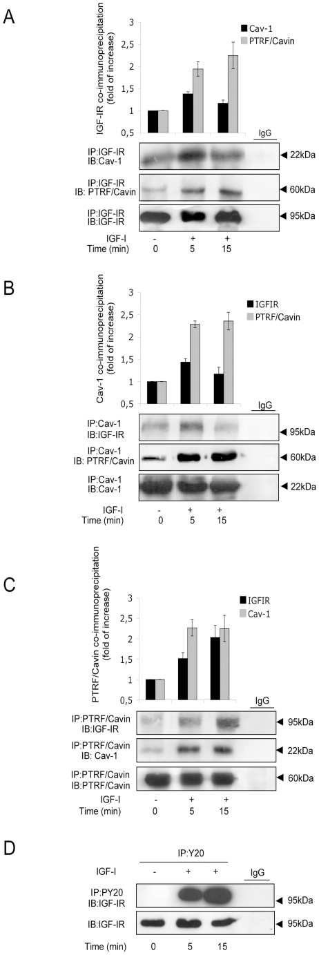
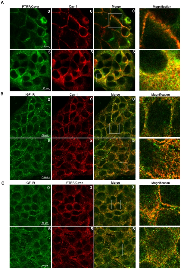
Background: Insulin-like growth factor-I receptor (IGF-IR) is a tyrosine kinase receptor (RTK) associated with caveolae, invaginations of the plasma membrane that regulate vesicular transport, endocytosis and intracellular signaling. IGF-IR internalization represents a key mechanism of down-modulation of receptors number on plasma membrane. IGF-IR interacts directly with Caveolin-1 (Cav-1), the most relevant protein of caveolae. Recently it has been demonstrated that the Polymerase I and Transcript Release Factor I (PTRF/Cavin) is required for caveolae biogenesis and function. The role of Cav-1 and PTRF/Cavin in IGF-IR internalization is still to be clarified.

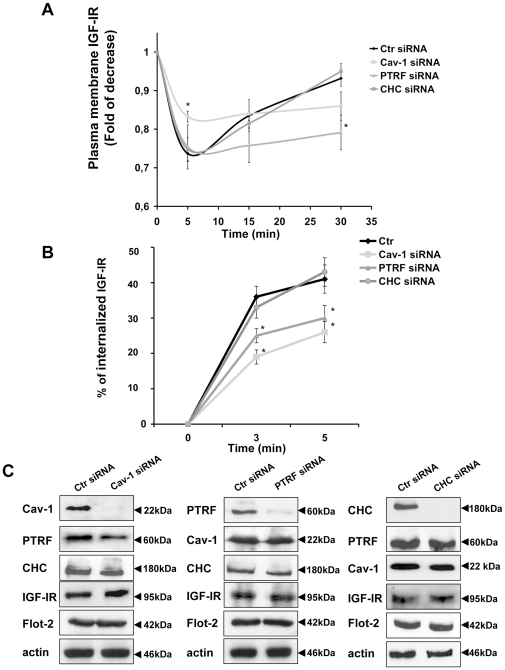
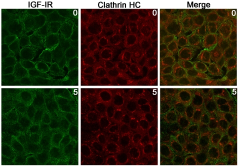
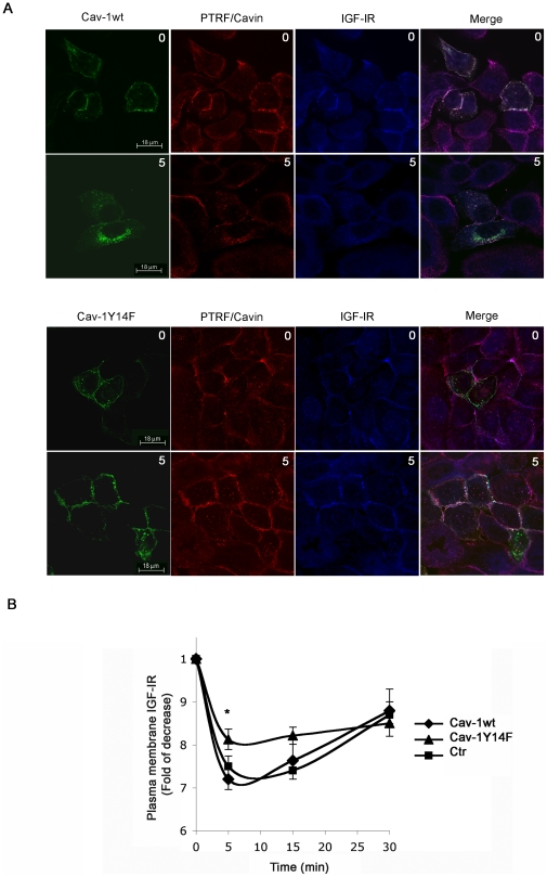
Methodology/principal findings: We have investigated the interaction of IGF-IR with Cav-1 and PTRF/Cavin in the presence of IGF1in human Hacat cells. We show that IGF-IR internalization triggers Cav-1 and PTRF/Cavin translocation from plasma membrane to cytosol and increases IGF-IR interaction with these proteins. In fact, Cav-1 and PTRF/Cavin co-immunoprecipitate with IGF-IR during receptor internalization. We found a different time course of co-immunoprecipitation between IGF-IR and Cav-1 compared to IGF-IR and PTRF/Cavin. Cav-1 and PTRF/Cavin silencing by siRNA differently affect surface IGF-IR levels following IGF1 treatment: Cav-1 and PTRF/Cavin silencing significantly affect IGF-IR rate of internalization, while PTRF/Cavin silencing also decreases IGF-IR plasma membrane recovery. Since Cav-1 phosphorylation could have a role in IGF-IR internalization, the mutant Cav-1Y14F lacking Tyr14 was transfected. Cav-1Y14F transfected cells showed a reduced internalization of IGF-IR compared with cells expressing wild type Cav-1. Receptor internalization was not impaired by Clathrin silencing. These findings support a critical role of caveolae in IGF-IR intracellular traveling.

Conclusions/significance: These data indicate that Caveolae play a role in IGF-IR internalization. Based on these findings, Cav-1 and PTRF/Cavin could represent two relevant and distinct targets to modulate IGF-IR function.


Conflict of interest statement

Competing Interests: The authors have declared that no competing interests exist.

Figures

Figure 1
Figure 1. Cav-1 and PTRF/Cavin co-immunoprecipitation with IGF-IR.
Serum starved HaCat cells were stimulated with IGF110 nM for the indicated times and then lysed. Equal amount of cell lysates were immunoprecipitated (IP) and immunoblotted (IB) with the indicated antibodies. The graphs represent quantification of co-immunoprecipitation experiments following densitometric analysis of bands and are expressed as fold of increase. Data shown are representative of three independent experiments and are expressed as the mean ± SD.
Figure 2
Figure 2. IGF1induces PTRF/Cavin and Cav-1 internalization.
HaCat cells were treated with IGF1 (10 nM) for 5 min, fixed in 4% formaldehyde, permeabilized with Methanol at −20°C, labeled with (A) a rabbit anti-PTRF/Cavin (red) and a mouse anti-Cav-1 (green), or (B) a mouse anti-IGF-IR (green) and a rabbit anti-Cav-1 antibody (red), or alternatively (C) with a mouse anti-IGF-IR (green) and a rabbit anti-PTRF/Cavin (red) antibody and imaged by confocal immunofluorescence microscopy. Column 4 shows magnified fields indicated by squares in column 3. Merged fields show co-localization (yellow) respectively of PTRF/Cavin and Cav-1 (A), IGFIR and Cav-1 (B) and of IGF-IR and PTRF (C).
Figure 3
Figure 3. Cav-1 and PTRF/Cavin are required for IGF-IR internalization and plasma membrane recovery.
HaCat cells were transfected with siRNA for Cav-1 (Cav-1-siRNA), for PTRF/Cavin (PTRF/Cavin-siRNA), for Clathrin Heavy Chain (Clathrin HC-siRNA) and with scrambled control siRNA (Ctr-siRNA) as described in materials and methods. (A) 72 hours after transfection, serum-starved cells were treated with IGF110 nM for the indicated times, trypsined, washed, blocked and incubated with a mouse PE-conjugated IGF-IR antibody. PE-conjugated IGF-IR labeled cells were analyzed by flow-cytometry to measure plasma membrane IGF-IR expression as described in Materials and Methods. (B) Ctr-siRNA, Cav-1-siRNA, PTRF/Cavin-siRNA and Clathrin HC-siRNA HaCat cells were serum-starved and subjected to a biotinylation based endocytic assay with NH-SS-biotin at 4°C (see Materials and Methods). The cells were then warmed at 37°C with medium containing IGF110 nM to allow IGF-IR internalization. Glutathione was used to reduce the proteins not internalized from the plasma membrane. IGF-IR was immunoprecipitated with IGF-IR antibody and the internalized IGF-IR was revealed by Western Blot with a Streptavidin-HRP antibody. Data were quantified using NIH-Image and plotted in the graph. The amount of biotinylated internalized IGF-IR was expressed as a percentage of the amount of IGF-IR on the surface at 4°C which we set as 100%. (C) 72 hours from the transfection serum-starved cells were lysed and equal amount of Ctr-siRNA and Cav-1-siRNA or Ctr-siRNA and PTRF/Cavin-siRNA and Clathrin HC-siRNA cell lysates were separated on SDS–PAGE, transferred on nitrocellulose and blotted with an antibody directed against Cav-1, PTRF/Cavin, Clathrin HC, IGF-IR, Flotillin-2 and actin proteins. Data are expressed as the mean ± SD. Statistical analysis was performed using Student's t test. *p<0.05.
Figure 4
Figure 4. IGF1 does not Induces IGF-IR and Clathrin Heavy Chain co-localization.
HaCat cells were treated with IGF1(10 nM) for 5 min, fixed in 4% formaldehyde, permeabilized with Methanol at −20°C, labeled with a rabbit anti-Clathrin Heavy Chain (red) and a mouse anti-IGF-IR (green), antibody and imaged by confocal immunofluorescence microscopy.
Figure 5
Figure 5. Expression of CavY14F mutant decreases IGF-IR internalization in IGF1 stimulated cells.
(A) Hacat cells were transiently transfected with pEGFPN1 Cav-1-wt and pEGFN1 Cav-1Y14F plasmids. 48 hours after transfection serum starved HaCat cells were stimulated with IGF1 10 nM for 5 min, fixed in 4% formaldehyde, permeabilized with Methanol at −20°C, labeled with a rabbit anti-PTRF/Cavin (red) and a mouse anti-IGF-IR (blue) imaged by confocal immunofluorescence microscopy. Merged fields show co-localization (white) of Cav-1wt, PTRF/Cavin, IGF-IR (upper pannels) and Cav-1Y14F, PTRF/Cavin and IGF-IR (bottom panels). (B) 48 hours from the transfection, serum-starved cells were treated with IGF110 nM for the indicated times, trypsined, washed, blocked and incubated with a mouse PE-conjugated IGF-IR.antibody. PE-conjugated IGF-IR labeled cells were analyzed by flow-cytometry to measure plasma membrane IGF-IR expression as described in Materials and Methods. Data are expressed as the mean ± SD. Statistical analysis was performed using Student's t test. *p<0.05.

References

    1. Werner H, Bruchim I. The insulin-like growth factor-I receptor as an oncogene. Arch Physiol Biochem. 2009;115:58–71. - PubMed
    1. Salani B, Briatore L, Garibaldi S, Cordera R, Maggi D. Caveolin-1 down regulation inhibits IGF-IR signal transduction in H9C2 rat cardio myoblasts. Endocrinology 2007 - PubMed
    1. Matthews LC, Taggart MJ, Westwood M. Effect of cholesterol depletion on mitogenesis and survival: the role of caveolar and noncaveolar domains in insulin-like growth factor-mediated cellular function. Endocrinology. 2005;146:5463–5473. - PubMed
    1. Baserga R. The IGF-I receptor in cancer research. Exp Cell Res. 1999;253:1–6. - PubMed
    1. Zapf A, Hsu D, Olefsky JM. Comparison of the intracellular itineraries of insulin-like growth factor-I and insulin and their receptors in Rat-1 fibroblasts. Endocrinology. 1994;134:2445–2452. - PubMed

Publication types

MeSH terms