. 2010 Nov 30;8(11):e1000547.
doi: 10.1371/journal.pbio.1000547.

The function and three-dimensional structure of a thromboxane A2/cysteinyl leukotriene-binding protein from the saliva of a mosquito vector of the malaria parasite

Affiliations

The function and three-dimensional structure of a thromboxane A2/cysteinyl leukotriene-binding protein from the saliva of a mosquito vector of the malaria parasite

Patricia H Alvarenga et al. PLoS Biol. .

Abstract

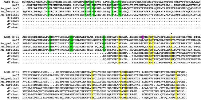
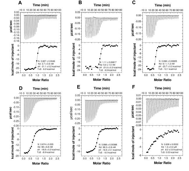
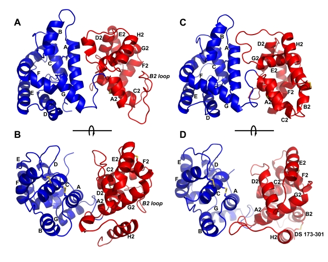
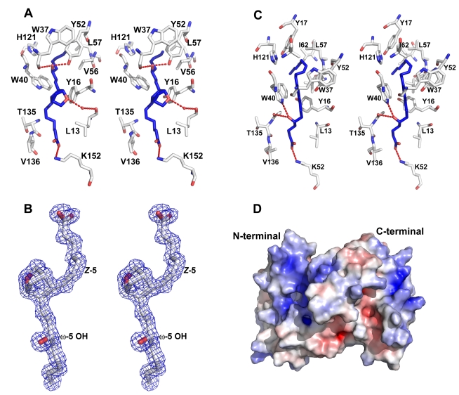
The highly expressed D7 protein family of mosquito saliva has previously been shown to act as an anti-inflammatory mediator by binding host biogenic amines and cysteinyl leukotrienes (CysLTs). In this study we demonstrate that AnSt-D7L1, a two-domain member of this group from Anopheles stephensi, retains the CysLT binding function seen in the homolog AeD7 from Aedes aegypti but has lost the ability to bind biogenic amines. Unlike any previously characterized members of the D7 family, AnSt-D7L1 has acquired the important function of binding thromboxane A(2) (TXA(2)) and its analogs with high affinity. When administered to tissue preparations, AnSt-D7L1 abrogated Leukotriene C(4) (LTC(4))-induced contraction of guinea pig ileum and contraction of rat aorta by the TXA(2) analog U46619. The protein also inhibited platelet aggregation induced by both collagen and U46619 when administered to stirred platelets. The crystal structure of AnSt-D7L1 contains two OBP-like domains and has a structure similar to AeD7. In AnSt-D7L1, the binding pocket of the C-terminal domain has been rearranged relative to AeD7, making the protein unable to bind biogenic amines. Structures of the ligand complexes show that CysLTs and TXA(2) analogs both bind in the same hydrophobic pocket of the N-terminal domain. The TXA(2) analog U46619 is stabilized by hydrogen bonding interactions of the ω-5 hydroxyl group with the phenolic hydroxyl group of Tyr 52. LTC(4) and occupies a very similar position to LTE(4) in the previously determined structure of its complex with AeD7. As yet, it is not known what, if any, new function has been acquired by the rearranged C-terminal domain. This article presents, to our knowledge, the first structural characterization of a protein from mosquito saliva that inhibits collagen mediated platelet activation.


Conflict of interest statement

The authors have declared that no competing interests exist.

Figures

Figure 1
Figure 1. Amino acid sequence alignment of D7 proteins from Anopheles sp. and Ae. aegypti.
The alignment shows sequences of the two-domain D7 forms from An. stephensi (AnSt-D7L1), An. gambiae, An. funestus, and An. darlingi in comparison to the two-domain protein from Aedes aegypti (AeD7). Also shown are the four single-domain D7 proteins (D7r1–4) from An. gambiae. Conserved residues contained in the ligand binding pocket of the N-terminal domain are highlighted in green, while those in the biogenic amine binding pocket of AeD7 and Anopheles single-domain D7s are highlighted in yellow. In the single-domain proteins and in the C-terminal domain of the two-domain proteins the conserved histidine residue (His 189 of AeD7) involved in biogenic amine binding is marked with an asterisk. Also highlighted in magenta is Trp 173 of AnSt-D7L1. In biogenic amine-binding forms, this residue is either leucine or valine.
Figure 2
Figure 2. Binding of CysLTs and TXA2 analogs by ITC.
Binding experiments were performed on a MicroCal VP-ITC instrument. For CysLT experiments the syringe contained a 20 µM ligand solution, and the AnSt-D7L1 concentration in the cell was 2 µM. For the TXA2 analog experiments the ligand solution used to fill the syringe was 40 µM and the AnSt-D7L1 concentration in the cell was 4 µM. Assays were performed at 35°C with successive 10 µL injections. The upper curve in each panel shows the measured heats for each injection, while the lower plot shows the enthalpies for each injection along with the fit to a single binding site model used to estimate the dissociation constant (Kd), enthalpy of binding (ΔH), entropy of binding (TΔS), and the binding stoichiometry (N). Panels: (A) LTC4, (B) LTD4, (C) LTE4, (D) U46619, (E) carbocyclic TXA2, (F) TXB2.
Figure 3
Figure 3. Binding of AnSt-D7L1 to prostaglandins and prostaglandins analogues by ITC.
Binding experiments were performed on a MicroCal VP-ITC instrument. For these experiments, the ligand solution used to fill the syringe was 80 µM and AnSt-D7L1 concentration in the calorimeter cell was 8 µM. Assays were performed at 35°C with successive 10 µL injections. Upper curve in each panel shows the heat for each injection; lower part shows the enthalpies for each injection and data were fit to a single binding site model used to calculate the binding stoichiometry (N), dissociation constant (Kd), enthalpy of binding (ΔH), and entropy of binding (TΔS). Panels: (A) PGD2, (B) PGE2, (C) PGF, (D) U51605.
Figure 4
Figure 4. Smooth muscle contraction bioassay.
(A) Guinea pig ileum samples were pre-incubated with buffer (solid line) or 1 µM AnSt-D7L1 (dotted line) and contraction was induced by 0.1 µM LTC4 (L), as indicated by the far left hand arrow, in both control and experimental preparations. Since in the presence of AnSt-D7L1 no contraction was observed after first LTC4 addition, further successive additions of 0.1 µM LTC4 were made only to this preparation (dotted line), as indicated by additional arrows. (B) Rat aorta samples were pre-incubated with buffer (solid line) or AnSt-D7L1 (dotted line) and the effect of addition of 0.1 µM U46619 (U) was recorded. Since there was no contraction in the presence of the AnSt-D7L1 after the first addition, further additions of 0.1 µM U46619 were made only to the preparation incubated with protein, as indicated by additional arrows. (C) The ability of AnSt-D7L1 to reverse contraction induced by U46619 was tested by pre-contacting rat aorta samples with 1 µM U46619 (as indicated by the letter “U”). After the plateau was achieved, in the experimental sample (dotted line) 0.1 µM of AnSt-D7L1 was added (indicated by D7 in the figure), which caused relaxation of the sample. In order to show that the muscle was still functional, 3 µM phenylephrine was added (indicated by PE). A control (solid line) experiment was performed without addition of protein or PE in order to show that in the absence of AnSt-D7L1 contraction lasts throughout the experiment.
Figure 5
Figure 5. Effect of AnSt-D7L1 on platelet aggregation promoted by different agonists.
Platelet-rich human plasma was incubated with different concentrations of AnSt-D7L1 for 1 min prior to the addition of agonists as indicated. Aggregation was measured at 37°C on an aggregometer. Unless otherwise stated protein concentration (µM) is shown under respective curves. (A) Concentration dependence of AnSt-D7L1 inhibition of collagen-mediated platelet aggregation at low concentration of collagen. (B) Lack of inhibition of collagen-mediated platelet aggregation by AnSt-D7L1 at high concentration of collagen. (C) Lack of inhibition of convulxin-mediated platelet aggregation by AnSt-D7L1. (D) ADP-mediated platelet aggregation in the absence of any protein (control), in the presence of 3 µM AnSt-D7L1, or in the presence of 3 µM RPAI1, which is known to bind ADP. (E) Lack of effect of AnSt-D7L1 on PMA-induced platelet aggregation. (F) Lack of effect of AnSt-D7L1 on ristocetin-induced platelet aggregation.
Figure 6
Figure 6. AnSt-D7L1 affects platelet aggregation promoted by TXA2.
Platelet-rich human plasma was incubated with different concentrations of AnSt-D7L1 or buffer, as indicated, for 1 min prior to the addition of agonists indicated in each panel. (A) Aggregation mediated by collagen in the presence of TXA2 antagonists. The effect of 3 µM AnSt-D7L1 on collagen-induced aggregation was similar to the effect promoted by a thromboxane receptor (TP) antagonist (SQ 29,548) or by pre-incubating platelets with indomethacin, a general cyclooxygenase inhibitor. The curve marked control shows the normal response of platelets to collagen. (B) AnSt-D7L1 has a dose dependent effect on U46619-induced platelet aggregation. Protein concentrations are shown on the bottom of each curve. The last curve in the panel shows the effect of 1 µM SQ 29,548 in the absence of AnSt-D7L1. (C) AnSt-D7L1 (3 µM) affects AA-induced platelet aggregation. A similar effect is observed when platelets are pre-incubated with indomethacin in the absence of protein, inhibiting TXA2 production. Respective treatments are indicated in the bottom of each curve.
Figure 7
Figure 7. Ribbon diagram of the AnSt-D7L1 structure and comparison with AeD7.
Structures of AnSt-D7L1 and AeD7 have the N-terminal domain colored blue and the C-terminal domain colored red. Cysteine residues forming disulfide bonds are shown in stick representation and colored yellow. (A) and (B) are two views of AnSt-D7L1 related by an approximately 90° rotation around the axis shown. (C) and (D) are two views of AeD7 from Ae. aegypti oriented in the same manner as (A) and (B). The helices are labeled A–G in the N-terminal domain and A2–H2 in the C-terminal domain. Helix B2 is missing in AnSt-D7L1, but the labeling of the other C-terminal domain helices corresponds to AeD7. The label “B2 loop” in (A) and (B) indicates the portion of the structure corresponding to α-helix B2 in AeD7. The label “DS 173–301” marks the position of the disulfide bond linking α-helix H2 with α-helix B2 in AeD7. The cysteine residues forming this bond are not present in AnSt-D7L1 (see panel B for comparison).
Figure 8
Figure 8. The AnSt-D7L1 N-terminal domain ligand binding site.
(A) Stereoview of the binding interactions of U46619 at the ligand binding site. In the ligand, carbon atoms are colored blue and oxygen atoms red. Protein residues have carbon colored white, oxygen red, and nitrogen blue. Hydrogen bonds are shown as dashed red lines. (B) Stereoview of the 2Fo-Fc electron density map covering the U46619 ligand at the AnSt-D7L1 binding site contoured at 1.0 σ. In the ligand, carbon atoms are colored blue and oxygen atoms red. The “cis” unsaturation at position 5 and the hydroxyl group at the ω-5 position are labeled. (C) Stereoview of binding interactions of LTC4 in the AnSt-D7L1 N-terminal domain ligand binding pocket. Since the peptide moiety of the ligand is not visible in the crystal structure, only the fatty acid portion (5(S)-hydroxy- (E,E,Z,Z)-7, 9, 11, 14-eicosatetraenoic acid) of the molecule is shown. In the ligand, carbon atoms are shown in blue and oxygen atoms in red. Protein atoms are colored as in (A). Hydrogen bonds are shown as red dashed lines. (D) The solvent-accessible surface of AnSt-D7L1 is colored by electrostatic potential. The N- and C-terminal domains are labeled. Note the concentration of positive electrostatic potential surrounding the binding site for U46619 and CysLTs.
Figure 9
Figure 9. Detail of the C-terminal domain structure of AnSt-D7L1 and AeD7.
(A) Stereoview of the region from the C-terminal domain of AnSt-D7L1 corresponding to the biogenic amine binding site of AeD7. Carbon atoms are colored in white, oxygen in red, nitrogen in blue. Helical segments are shown in ribbon format and hydrogen bonds are shown as dashed red lines. (B) Stereoview of the biogenic amine binding pocket (including a bound norepinephrine molecule labeled NE) of AeD7 shown from approximately the same view as AnSt-D7L1 in panel (A). Helical segment B2 is labeled to highlight how the conformational difference in this portion of the structure affects the positioning of residues in the ligand-binding pocket.

References

    1. Ribeiro J. M. Blood-feeding arthropods: live syringes or invertebrate pharmacologists? Infect Agents Dis. 1995;4:143–152. - PubMed
    1. Ribeiro J. M, Francischetti I. M. Role of arthropod saliva in blood feeding: sialome and post-sialome perspectives. Annu Rev Entomol. 2003;48:73–88. - PubMed
    1. Champagne D. E. Antihemostatic molecules from saliva of blood-feeding arthropods. Pathophysiol Haemost Thromb. 2005;34:221–227. - PubMed
    1. Valenzuela J. G. High-throughput approaches to study salivary proteins and genes from vectors of disease. Insect Biochem Mol Biol. 2002;32:1199–1209. - PubMed
    1. Titus R. G, Ribeiro J. M. Salivary gland lysates from the sand fly Lutzomyia longipalpis enhance Leishmania infectivity. Science. 1988;239:1306–1308. - PubMed

Publication types

MeSH terms