. 2010 Nov 30;5(11):e14127.
doi: 10.1371/journal.pone.0014127.

Independent interactions of phosphorylated β-catenin with E-cadherin at cell-cell contacts and APC at cell protrusions

Affiliations

Independent interactions of phosphorylated β-catenin with E-cadherin at cell-cell contacts and APC at cell protrusions

Maree C Faux et al. PLoS One. .

Abstract

Background: The APC tumour suppressor functions in several cellular processes including the regulation of β-catenin in Wnt signalling and in cell adhesion and migration.

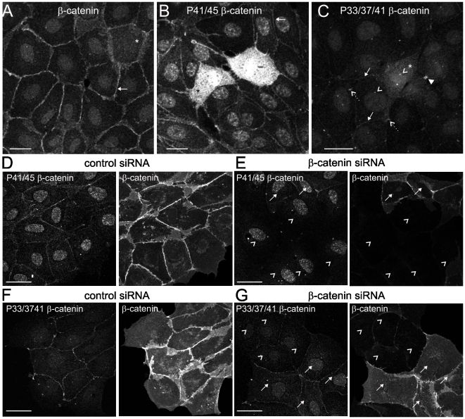
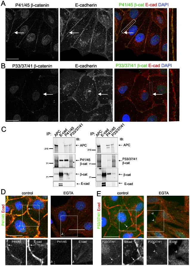
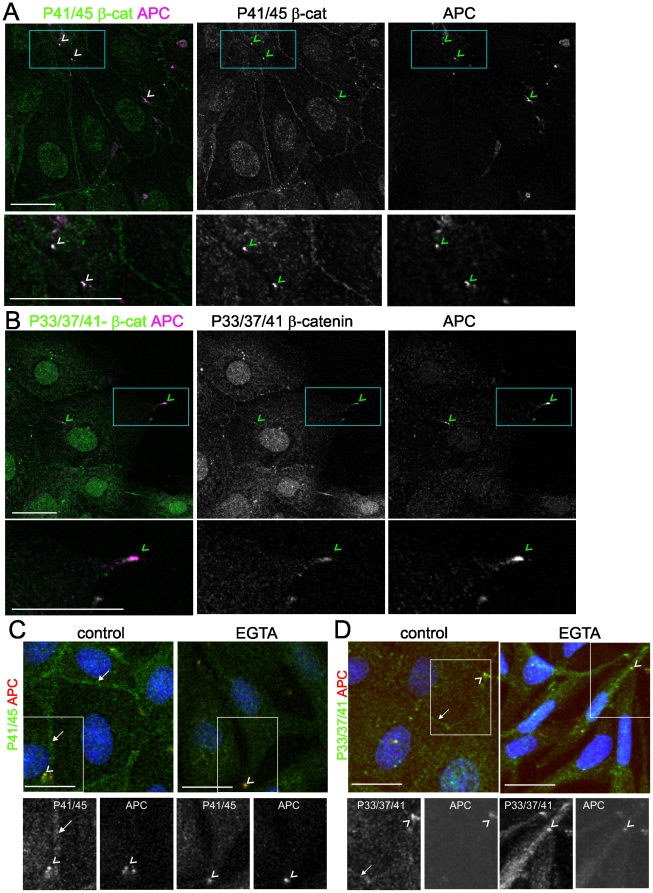
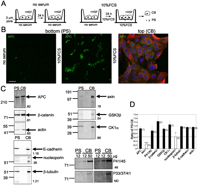
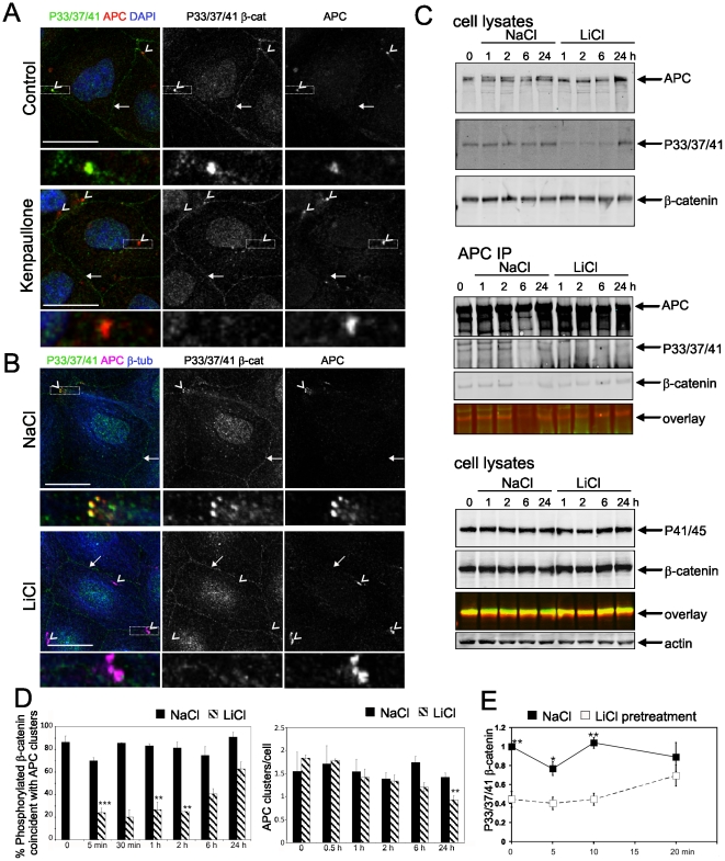
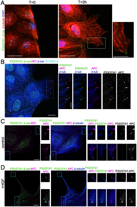
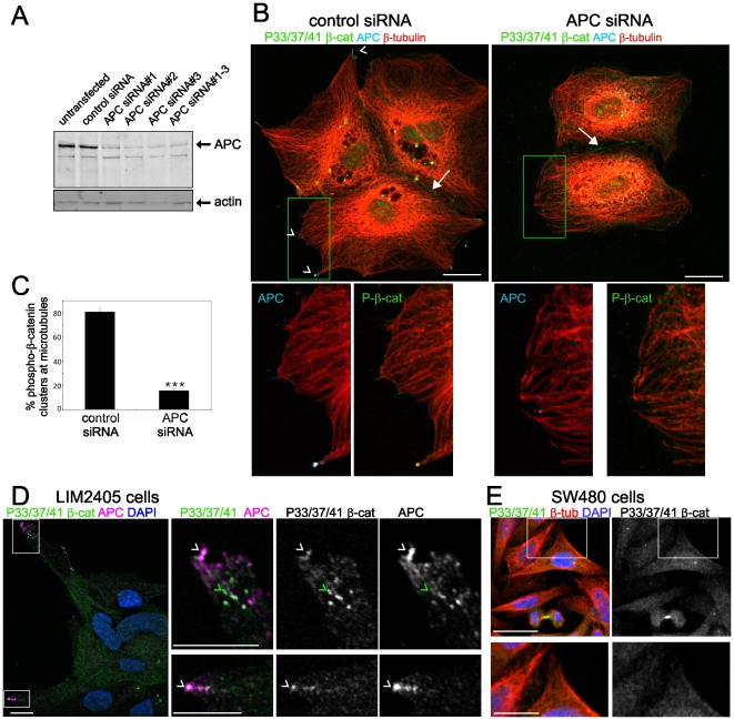
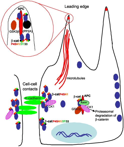
Findings: In this study, we establish that in epithelial cells N-terminally phosphorylated β-catenin specifically localises to several subcellular sites including cell-cell contacts and the ends of cell protrusions. N-terminally phosphorylated β-catenin associates with E-cadherin at adherens junctions and with APC in cell protrusions. We isolated APC-rich protrusions from stimulated cells and detected β-catenin, GSK3β and CK1α, but not axin. The APC/phospho-β-catenin complex in cell protrusions appears to be distinct from the APC/axin/β-catenin destruction complex. GSK3β phosphorylates the APC-associated population of β-catenin, but not the cell junction population. β-catenin associated with APC is rapidly phosphorylated and dephosphorylated. HGF and wound-induced cell migration promote the localised accumulation of APC and phosphorylated β-catenin at the leading edge of migrating cells. APC siRNA and analysis of colon cancer cell lines show that functional APC is required for localised phospho-β-catenin accumulation in cell protrusions.

Conclusions: We conclude that N-terminal phosphorylation of β-catenin does not necessarily lead to its degradation but instead marks distinct functions, such as cell migration and/or adhesion processes. Localised regulation of APC-phospho-β-catenin complexes may contribute to the tumour suppressor activity of APC.


Conflict of interest statement

Competing Interests: The authors have declared that no competing interests exist.

Figures

Figure 1
Figure 1. Populations of phosphorylated β-catenin in epithelial cells.
(A) β-catenin is localised to cell-cell contacts (arrow). Mitotic cell is indicated by *. (B) Phospho-β-catenin pT41/pT45 (P41/45) is localised to the cell membrane (arrow) and mitotic cell cytoplasm (*). (C) Phospho β-catenin pS33/pS37/pT41 (P33/37/41) is localised to the cell membrane (arrows), in discrete clusters (dashed arrows) and the mitotic cell cytoplasm (*). The staining at centrosomes (open arrowheads) and mid-body bridges (filled arrowhead) is indicated. (D–G) MDCK cells were treated with siRNA directed against β-catenin or a control sequence. Cells were fixed 72 h after siRNA transfection and immunostained with antibodies to P41/45 β-catenin and β-catenin (D and E) and P33/37/41 β-catenin and β-catenin (F and G). Fields of cells without knockdown (arrows) are shown adjacent to β-catenin depleted cells (arrowheads) for comparison. Scale bars 20 µm.
Figure 2
Figure 2. Phosphorylated β-catenin associates with E-cadherin at cell-cell adherens junctions.
(A) P41/45-β-catenin is localised to cell-cell contacts with E-cadherin. (B) P33/37/41-β-catenin is localised to the cell periphery and shows partial overlap with E-cadherin. Cell contacts are indicated by arrows. Inset, enlarged view of overlapping phospho-β-catenin (green) and E-cadherin (red). P41/45-β-catenin (A) shows more prominent cell contact staining and overlap with E-cadherin than P33/37/41-β-catenin (B). (C) Co-precipitation of phospho-β-catenin with E-cadherin and APC in MDCK cells. APC, E-cadherin, P41/45- and P33/37/41-β-catenin immunoprecipitates were immuno-blotted with the indicated antibodies. * denotes non-specific band. (D and E) Peripheral phospho-β-catenin is regulated by Ca2+-dependent cell-cell contact. Cell contact associated E-cadherin and phospho-β-catenin staining is intact in control cells (arrows), but disrupted in treated cells where phospho β-catenin clusters remain (arrowheads). Phospho-β-catenin (green), E-cadherin (red), nuclei (DAPI) blue. Shown are representatives of at least three different experiments; scale bars 20 µm.
Figure 3
Figure 3. Phosphorylated β-catenin associates with APC at the ends of microtubules.
(A and B) Phosphorylated β-catenin is localised in clusters with APC in MDCK cells. Coincident APC (magenta) and phospho-β-catenin (green) is indicated with arrowheads. (C and D) Disruption of Ca2+-dependent cell-cell contact does not alter phospho-β-catenin coincident with APC in clusters at cell extensions (arrowheads). Junctional phospho-β-catenin staining is intact in control cells (arrows). Phospho-β-catenin (green), APC (red), nuclei (DAPI) blue. Shown are representatives of at least three different experiments; scale bars 20 µm.
Figure 4
Figure 4. Isolation of APC from protruding pseduopodia.
(A) Schematic diagram depicting strategy for isolation of pseudopodia. PS pseudopodia, CB cell bodies. (B) MDCK cells on 3.0 µm pore filters were induced to extend pseudopodial protrusions. Cells were fixed and either cell bodies were removed leaving pseudopodia, bottom (PS), or pseduopodia were removed leaving cell bodies, top (CB). Filters were immunostained for APC (green) and β-tubulin (red) and costained with DAPI (blue).Cells protrusions contain APC. DAPI staining confirms there are no nuclei on the lower surface, bottom (PS). Scale bar, 20 µm. (C) Isolation of APC and β-catenin, but not axin, from the pseudopodia (PS) fraction. Immunoblots of proteins isolated from the pseudopodia (PS) on the lower membrane surface or cell bodies (CB) on the upper membrane surface of 3.0 µm pore filters. Total protein loaded was 12 µg except for the P41/45 and P33/37/41 immunoblots where 50 µg CB was loaded in order to detect the proteins. The numbers to the side of blots are the normalised ratio of the amount of each protein in PS vs CB. Shown is representative from at least 3 independent experiments. (D) APC, but not axin, is present in the PS fraction. Proteins isolated from the PS or CB fractions were resolved by SDS-PAGE and immunoblotted as in (C). Shown is the average +/− SEM of the ratio of PS∶CB for the indicated protein from at least three separate experiments (n is indicated). ***P<0.002, *P<0.05 by Student's t-test.
Figure 5
Figure 5. Inhibition of GSK3β results in loss of P33/37/41-β-catenin associated with APC but not other sub-populations.
(A) Kenpaullone (2 nM, 30 min) and (B) LiCl (30 mM, 5 min) treatment of MDCK cells results in loss of P33/37/41-β-catenin coincident with APC. APC clusters (arrowheads) are coincident with P33/37/41-β-catenin in control but not kenpaullone or LiCl treated cells; intact peripheral staining is indicated by arrows. Scale bars 20 µm. (C) LiCl treatment results in reduced P33/37/41-β-catenin in total cell lysates and associated with APC, but not P41/45-β-catenin. Lysates from MDCK cells treated with 30 mM NaCl or LiCl for T0, 1, 2, 6 and 24 h were immunoblotted or immunoprecipitated with APC antibodies and immunoblotted as indicated. Overlay refers to the merge of P33/37/41 or P41/45-anti-rabbit-IR800 and β-catenin-anti-mouse-IR680 channels. (D) Analysis of coincident P33/37/41-β-catenin and APC clusters following GSK3β inhibition. MDCK cells were treated with 30 mM NaCl or LiCl for the indicated times, fixed and immunostained. Cells were scored for coincident P33/37/41-β-catenin and APC clusters (left) and number of APC clusters/cell (right). Shown is average +/− SEM from three (T0, 5 min, 1, 2 6 and 24 h) or two (T30 min) independent experiments, >100 cells per treatment group; ***P<0.001, **P<0.01 by Student's t-test. (E) Removal of GSK3β inhibition allows re-phosphorylation of β-catenin. MDCK cells pretreated with 30 mM NaCl or LiCl for 30 min, were incubated in DME+10%FCS and harvested at T0, 5, 10 and 20 min. Lysates were immunoblotted with P33/37/41 β-catenin and actin antibodies. Shown is average +/− SEM from four independent experiments for normalised densitometry values, *P<0.05, **P<0.01 by Student's t-test. A representative blot is shown in Fig S10A.
Figure 6
Figure 6. Phospho-β-catenin and APC are localised to the ends of microtubules in migrating cells.
(A) P33/37/41-β-catenin in clusters at the wound edge. Representative images of cells fixed at T = 0 (left) and 2 hours after wounding. P33/37/41-β-catenin (green), β-tubulin (red), DAPI (blue). Scale 20 µm; inset 10 µm. (B) P33/37/41-β-catenin and APC in migrating MDCK cells. Merged image shows coincident (white) P33/37/41-β-catenin (green) and APC (magenta) at the ends of microtubules (β-tubulin, blue). Nuclei were visualised with TO-PRO-3 iodide staining (cyan). Scale 10 µm. Insets show enlarged view of the leading edge of migrating cells. Scale bar 5 µm. (C and D) MDCK cells were serum starved and untreated (C) or stimulated with 2 ng/ml HGF for 6 h (D) and immunostained with antibodies to P33/37/41-β-catenin (green), APC (magenta) and β-tubulin (blue). Scale 10 µm. Insets show clusters at microtubule ends that are highly concentrated in HGF treated cells. Scale bar 5 µm.
Figure 7
Figure 7. APC is required for phospho-β-catenin localisation to the ends of microtubules.
(A) Depletion of cellular APC by siRNA. APC levels following siRNA transfection were assessed by immunoblot analysis in cell lysates. Actin was used as a loading control. (B) MDCK cells transfected with control and APC siRNA were immunostained for P33/37/41-β-catenin (green), APC (cyan) and β-tubulin (red) with APC/β-tubulin and P33/37/41-β-catenin/β-tubulin enlarged in insets shown below. Arrowheads indicate clusters of phospho-β-catenin at microtubule ends coincident with APC in control but not APC depleted cells. Scale bars 20 µm. (C) Quantification of phospho-β-catenin clusters (%) at microtubule protrusions in control and APC siRNA treated cells. Shown is average +/− SD from 3 independent experiments, >70 microtubule protrusions per treatment group. ***P<0.001 by Student's t-test. (D) LIM2405 colon cancer cells containing a wt APC allele demonstrate concentrations of P33/37/41-β-catenin (green) at cell extensions that partially overlap with APC (magenta) (white arrowheads indicate overlap with APC, green arrowheads indicate concentrations of P33/37/41-β-catenin that do not overlap with APC). Nuclei were visualised with DAPI; scale bars 10 µm. (E) P33/37/41-β-catenin is diffusely cytoplasmic in SW480 colon cancer cells and does not concentrate in clusters at microtubule ends. SW480 cells were immunostained with P33/37/41-β-catenin (green) and β-tubulin (red) and costained with DAPI to visualise nuclei, scale bar 20 µm. P33/37/41/β-tubulin and P33/37/41-β-catenin enlarged in insets are shown below, scale bar 10 µm.
Figure 8
Figure 8. Functionally distinct populations of N-terminally phosphorylated β-catenin.
P41/45-β-catenin associates with E-cadherin at cell-cell contacts. P33/37/41-β-catenin is also found at Ca2+-dependent cell contacts but is not detected in the complex with E-cadherin. P41/45- and P33/37/41-β-catenin associate with APC at the ends of microtubules at the leading edge of migrating cells. At the protrusions the phospho-β-catenin is rapidly phosphorylated and dephosphorylated. This complex is separate from the axin destruction complex that targets phospho-β-catenin for proteasomal degradation.

Similar articles

Cited by

References

    1. Cottrell S, Bicknell D, Kaklamanis L, Bodmer WF. Molecular analysis of APC mutations in familial adenomatous polyposis and sporadic colon carcinomas. Lancet. 1992;340:626–630. - PubMed
    1. Powell SM, Zilz N, Beazer-Barclay Y, Bryan TM, Hamilton SR, et al. APC mutations occur early during colorectal tumorigenesis. Nature. 1992;359:235–237. - PubMed
    1. Giles RH, van Es JH, Clevers H. Caught up in a Wnt storm: Wnt signaling in cancer. Biochim Biophys Acta. 2003;1653:1–24. - PubMed
    1. Huang H, He X. Wnt/beta-catenin signaling: new (and old) players and new insights. Curr Opin Cell Biol. 2008;20:119–125. - PMC - PubMed
    1. Aberle H, Bauer A, Stappert J, Kispert A, Kemler R. beta-catenin is a target for the ubiquitin-proteasome pathway. Embo J. 1997;16:3797–3804. - PMC - PubMed

Publication types

MeSH terms