. 2010 Nov 30;5(11):e14137.
doi: 10.1371/journal.pone.0014137.

Screening of human tumor antigens for CD4 T cell epitopes by combination of HLA-transgenic mice, recombinant adenovirus and antigen peptide libraries

Affiliations

Screening of human tumor antigens for CD4 T cell epitopes by combination of HLA-transgenic mice, recombinant adenovirus and antigen peptide libraries

Wolfram Osen et al. PLoS One. .

Abstract

Background: As tumor antigen-specific CD4+ T cells can mediate strong therapeutic anti-tumor responses in melanoma patients we set out to establish a comprehensive screening strategy for the identification of tumor-specific CD4+ T cell epitopes suitable for detection, isolation and expansion of tumor-reactive T cells from patients.

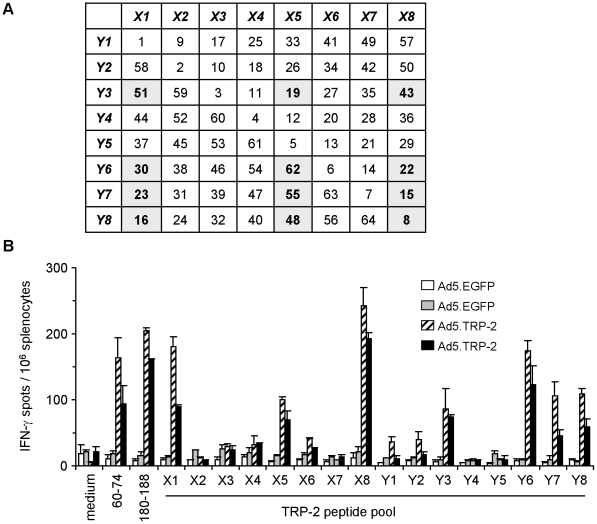
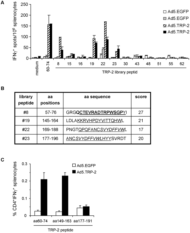
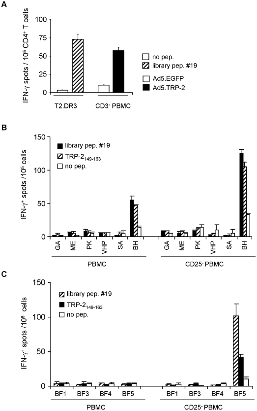
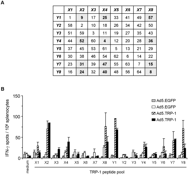
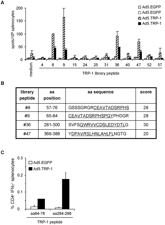
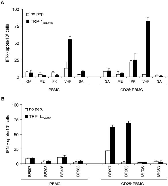
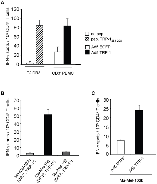
Methods and findings: To scan the human melanoma differentiation antigens TRP-1 and TRP-2 for HLA-DRB1*0301-restricted CD4+ T cell epitopes we applied the following methodology: Splenocytes of HLA-DRB1*0301-transgenic mice immunized with recombinant adenovirus encoding TRP-1 (Ad5.TRP-1) or TRP-2 (Ad5.TRP-2) were tested for their T cell reactivity against combinatorial TRP-1- and TRP-2-specific peptide libraries. CD4+ T cell epitopes thus identified were validated in the human system by stimulation of peripheral blood mononuclear cells (PBMC) from healthy donors and melanoma patients. Using this strategy we observed that recombinant Ad5 induced strong CD4+ T cell responses against the heterologous tumor antigens. In Ad5.TRP-2-immunized mice CD4+ T cell reactivity was detected against the known HLA-DRB1*0301-restricted TRP-2(60-74) epitope and against the new epitope TRP-2(149-163). Importantly, human T cells specifically recognizing target cells loaded with the TRP-2(149-163)-containing library peptide or infected with Ad5.TRP-2 were obtained from healthy individuals, and short term in vitro stimulation of PBMC revealed the presence of epitope-reactive CD4+ T cells in melanoma patients. Similarly, immunization of mice with Ad5.TRP-1 induced CD4+ T cell responses against TRP-1-derived peptides that turned out to be recognized also by human T cells, resulting in the identification of TRP-1(284-298) as a new HLA-DRB1*0301-restricted CD4+ T cell epitope.

Conclusions: Our screening approach identified new HLA-DRB1*0301-restricted CD4+ T cell epitopes derived from melanoma antigens. This strategy is generally applicable to target antigens of other tumor entities and to different HLA class II molecules even without prior characterization of their peptide binding motives.


Conflict of interest statement

Competing Interests: The authors have declared that no competing interests exist.

Figures

Figure 1
Figure 1. Combinatorial peptide library screening allows detection of individual library peptides containing TRP-2-specific T cell epitopes.
A, Composition of the TRP-2-specific library peptide pools X1 to X8 and Y1 to Y8 used for combinatorial screening of specific T cell responses ex vivo. Individual library peptides determined by combinatorial screening are highlighted. B, Spleen cells from HLA-DR3tg mice injected i.p. with 5×10⁸ pfu Ad5.TRP-2 or Ad5.EGFP (2 mice per group) were screened ex vivo by IFN-γ ELISpot assay for recognition of TRP-2-specific library peptide pools. T cell responses of two control mice (Ad5.EGFP) and two TRP-2-immunized mice (Ad5.TRP-2) are represented as individual columns in the diagram. Reactivity against two controls, the H2-Kb-restricted CD8+ T cell epitope TRP-2180-188 and the HLA-DRB1*0301-restricted CD4+ T cell epitope TRP-260–74 was tested in addition. Error bars show standard error of the mean. Experiments were performed four times, yielding similar results.
Figure 2
Figure 2. Phenotypic analysis of TRP-2-specific T cell responses induced in Ad5.TRP-2-immunized HLA-DR3tg mice.
A, Spleen cells from HLA-DR3tg mice injected i.p. with 5×10⁸ pfu Ad5.TRP-2 or Ad5.EGFP (2 mice per group) were screened ex vivo by IFN-γ ELISpot assay for reactivity against individual TRP-2-specific library peptides determined by combinatorial analysis. T cell responses of two control mice (Ad5.EGFP) and two TRP-2-immunized mice (Ad5.TRP-2) are represented as individual columns in the diagram. Error bars show standard error of the mean. Experiments were performed three times, yielding similar results. B, Selected TRP-2-derived library peptides #8, #19, #22 and #23 are indicated by amino acid (aa) positions and aa sequence. Peptides were analyzed in silico by the SYFPEITHI algorithm for the presence of predicted HLA-DRB*0301 binding sequences (underlined). Prediction scores for HLA-DRB1*0301-restricted epitopes are listed on the right. Known HLA-DRB1*0301-restricted CD4+ T cell epitopes are typed in bold and the H2-Kb-restricted CTL epitope TRP-2180–188 is written in italics. C, HLA-DR3tg mice received i.p. injections of 5×10⁸ pfu Ad5.TRP-2 or Ad5.EGFP (3 mice per group). Two weeks later spleen cells from infected mice were harvested and stimulated in vitro with the indicated peptides. After 6 days, splenocyte cultures were analyzed for the presence of peptide-reactive T cells by intracellular IFN-γ staining. Stained cells were analyzed by flow cytometry for the percentage of IFN-γ+ CD4+ T cells. Error bars show standard error of the mean of three immunized mice. Experiments were performed three times yielding similar results.
Figure 3
Figure 3. CD4+ T cells from melanoma patients respond to the HLA-DRB1*0301-restricted epitope TRP-2149–163.
A, PBMC from a HLA-DRB1*0301+ healthy donor were primed in vitro with autologous DC pulsed with library peptide #19. After two rounds of in vitro restimulation, CD4+ T cells were tested against T2.DR3 target cells pulsed with library peptide #19 and for recognition of autologous CD3+-depleted PBMC (CD3 PBMC) infected with Ad5.TRP-2 or with control virus. One representative experiment out of two is presented. B, Total PBMC (left panel) or CD25+-depleted PBMC (right panel) from six HLA-DRB1*03+ melanoma patients and C, four HLA-DRB1*0301+ healthy donors were incubated in vitro with library peptide #19. After 17 days, cells were tested by IFN-γ ELISpot assay for their reactivity against library peptide #19 or the epitope TRP-2149–163. All determinations were performed at least in duplicates. Data are presented as mean numbers of IFN-γ spots per 105 cells. Error bars show standard error of the mean.
Figure 4
Figure 4. Combinatorial peptide library screening allows detection of individual library peptides containing TRP-1-specific T cell epitopes.
A, Composition of the TRP-1-specific library peptide pools X1 to X8 and Y1 to Y8 used for combinatorial screening of specific T cell responses ex vivo. Individual peptides determined by combinatorial screening are highlighted. B, Spleen cells from HLA-DR3tg mice injected i.p. with 5×10⁸ pfu Ad5.TRP-1 or Ad5.EGFP (2 mice per group) were screened ex vivo by IFN-γ ELISpot assay for recognition of TRP-1-specific library peptide pools. T cell responses of two control mice (Ad5.EGFP) and two Ad5.TRP-1-immunized mice are represented as individual columns in the diagram. Error bars show standard error of the mean of duplicates. Experiments were performed four times, yielding similar results.
Figure 5
Figure 5. Phenotypic analysis of TRP-1-specific T cell responses induced in HLA-DR3tg mice upon immunization with Ad5.TRP-1.
A, Spleen cells from HLA-DR3tg mice injected i.p. with 5×10⁸ pfu Ad5.TRP-1 or Ad5.EGFP (2 mice per group) were screened ex vivo by IFN-γ ELISpot assay for reactivity against selected TRP-1-derived library peptides. T cell responses of two control mice (Ad5.EGFP) and two Ad5.TRP-1-immunized mice are represented as individual columns in the diagram. Error bars show standard error of the mean of duplicates. Experiments were performed three times, yielding similar results. B, Selected TRP-1-derived library peptides #8, #9, #36 and #47 are indicated by amino acid (aa) positions and aa sequence. Peptides were analyzed in silico by the SYFPEITHI algorithm for the presence of potential HLA-DRB*0301 binding sequences (underlined) . Prediction scores for HLA-DRB1*0301-restricted epitopes are listed on the right. The potential H2-restricted CTL epitope TRP-1374–382 is given in italics. C, Spleen cells of HLA-DR3tg mice injected i.p. with 5×10⁸ pfu Ad5.TRP-1 or Ad5.EGFP (3 mice per group) were analyzed for the presence of peptide-reactive T cells by intracellular IFN-γ staining after one round of in vitro re-stimulation with the indicated peptides. Note: The amino acid sequence of these peptides was shifted towards the N-terminus by one residue relative to the predicted epitope sequence. Stained cells were analyzed by flow cytometry for the percentage of IFN-γ+ CD4+ T cells. Error bars show standard error of the mean of three immunized mice. Experiments were performed twice yielding similar results.
Figure 6
Figure 6. Detection of TRP-1284–298 reactive CD4+ T cells in melanoma patients.
Total PBMC (left panel) or CD25+-depleted PBMC (right panel) from five HLA-DRB1*03+ melanoma patients (A) and four HLA-DRB1*0301+ healthy donors (B) were stimulated in vitro with peptide TRP-1284–298. After 25 days, PBMC were tested for their peptide reactivity by IFN-γ ELISpot assay. All determinations were performed at least in duplicates. The data are presented as mean numbers of IFN-γ spots per 105 cells. Error bars show standard error of the mean.
Figure 7
Figure 7. TRP-1284–298 represents a HLA-DRB1*0301-restricted CD4+ T cell epitope processed by human target cells.
PBMC from melanoma patient VHP were stimulated once with peptide TRP-1284–298 (10 µg/ml). After 25 days, selected CD4+ T cells were tested for their reactivity against different target cells by IFN-γ ELISpot assay. A, Recognition of T2.DR3 target cells pulsed with peptide TRP-1284–298 and autologous CD3+-depleted PBMC (CD3 PBMC) infected with Ad5.TRP-1 or with control virus is depicted. B, Reactivity of T cells against different melanoma cell lines Ma-Mel-103b (HLA-DRB1*0301+, TRP-1), Ma-Mel-108 (HLA-DRB1*0301+, TRP-1+), Ma-Mel-153 (HLA-DRB1*0301, TRP-1+) and C, against Ma-Mel-103b cells infected with Ad5.TRP-1 or control virus is presented. A–C, One representative out of two to three independent experiments is given. Error bars show standard error of the mean.

References

    1. Galon J, Costes A, Sanchez-Cabo F, Kirilovsky A, Mlecnik B, et al. Type, density, and location of immune cells within human colorectal tumors predict clinical outcome. Science. 2006;313:1960–1964. - PubMed
    1. Naito Y, Saito K, Shiiba K, Ohuchi A, Saigenji K, et al. CD8+ T Cells infiltrated within cancer cell nests as a prognostic factor in human colorectal cancer. Cancer Res. 1998;58:3491–3494. - PubMed
    1. Zhang L, Conejo-Garcia JR, Katsaros D, Gimotty PA, Massobrio M, et al. Intratumoral T Cells, recurrence, and survival in epithelial ovarian cancer. N Engl J Med. 2003;348:203–213. - PubMed
    1. Ferradini L, Mackensen A, Genevee C, Bosq J, Duvillard P, et al. Analysis of T cell receptor variability in tumor-infiltrating lymphocytes from a human regressive melanoma. Evidence for in situ T cell clonal expansion. J Clin Invest. 1993;91:1183–1190. - PMC - PubMed
    1. Yee C, Thompson JA, Byrd D, Riddell SR, Roche P, et al. Adoptive T cell therapy using antigen-specific CD8+ T cell clones for the treatment of patients with metastatic melanoma: in vivo persistence, migration, and antitumor effect of transferred T cells. Proc Natl Acad Sci U S A. 2002;99:16168–16173. - PMC - PubMed

Publication types

MeSH terms