. 2011 Mar;22(3):883-91.
doi: 10.1007/s00198-010-1499-4. Epub 2010 Dec 10.

Maternal vitamin D status affects bone growth in early childhood--a prospective cohort study

Affiliations

Maternal vitamin D status affects bone growth in early childhood--a prospective cohort study

H T Viljakainen et al. Osteoporos Int. 2011 Mar.

Abstract

In this prospective study, 87 children were followed up from birth to 14 months with data on maternal vitamin D status during the pregnancy. Postnatal vitamin D supplementation improved vitamin D status but only partly eliminated the differences in bone variables induced by maternal vitamin D status during the fetal period.

Introduction: Intrauterine nutritional deficits may have permanent consequences despite improved nutritional status postnatally. We evaluated the role of prenatal and postnatal vitamin D status on bone parameters in early infancy.

Methods: Eighty-seven children were followed from birth to 14 months. Background data were collected with a questionnaire and a 3-day food record. At 14 months bone variables were measured with peripheral computed tomography (pQCT) from the left tibia. Serum 25-OHD and bone turnover markers were determined. Findings were compared with maternal vitamin D status during pregnancy.

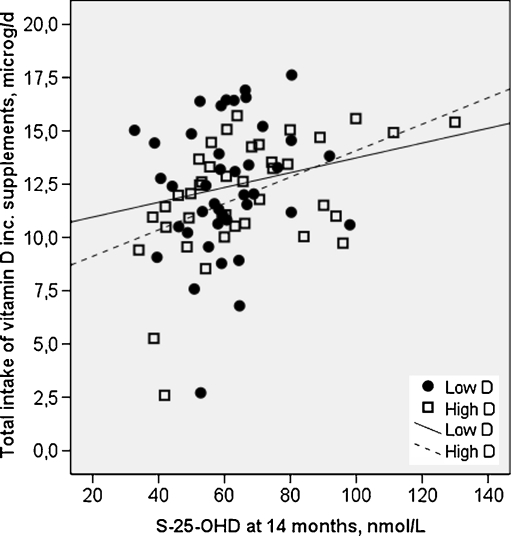
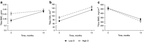
Results: The children were divided into two groups based on vitamin D status during pregnancy. Despite discrepant S-25-OHD at baseline (median 36.3 vs. 52.5 nmol/l, p < 0.001), the values at 14 months were similar (63 vs. 66 nmol/l, p = 0.58) in Low D and High D. Serum 25-OHD increased more in Low D (p < 0.001) despite similar total intake of vitamin D (mean 12.3 μg/day). In Low D, tibial bone mineral content (BMC) was lower at birth but BMC gain was greater (multivariate analysis of variance [MANOVA]; p = 0.032) resulting in similar BMC at 14 months in the two groups. In High D, tibial total bone cross-sectional area was higher at baseline; the difference persisted at 14 months (MANOVA; p = 0.068). Bone mineral density (BMD) and ΔBMD were similar in the two groups.

Conclusions: Postnatal vitamin D supplementation improved vitamin D status but only partly eliminated the differences in bone variables induced by maternal vitamin D status during the fetal period. Further attention should be paid to improving vitamin D status during pregnancy.


Figures

Fig. 1
Fig. 1
Total intake of vitamin D correlated positively with serum 25-OHD in High D (r = 0.505, p < 0.001), but not in Low D (r = 0.219, p = 0.168). Squares indicate High D and circles indicate Low D
Fig. 2
Fig. 2
BMC, CSA and BMD in study groups from baseline to 14 months. Increase in BMC from baseline to 14 months differed between the groups (repeated-measures MANOVA; p = 0.023) (a) due to higher baseline BMC in High D. No difference was observed in BMC between the groups at the 14-month visit. TB CSA from baseline to 14 months was significantly higher in High D than in Low D (repeated-measures MANOVA; p = 0.004) (b) due to the higher baseline CSA in High D. At 14 months, CSA remained 14.6 (SEM = 7.8) mm2 (MANOVA; p = 0.068) higher in High D. There was no difference between the groups in BMD during the 14 months (c) or in ΔBMD. Low D and High D groups are represented by circles and squares, respectively. Error bars represent SEMs
Fig. 3
Fig. 3
Concentrations of BALP and TRACP in study groups from baseline to 14 months. Low D and High D are represented by circles and squares, respectively. Error bars represent SEM. BALP increased from baseline (repeated-measures MANOVA; p < 0.001) (a) while TRACP concentration remained at the same level during the 14 months (b). There were no differences between the study groups

References

    1. Cooper C, Fall C, Egger P, Hobbs R, Eastell R, Barker D. Growth in infancy and bone mass in later life. Ann Rheum Dis. 1997;56:17–21. doi: 10.1136/ard.56.1.17. - DOI - PMC - PubMed
    1. Yarbrough DE, Barrett-Connor E, Morton DJ. Birth weight as a predictor of adult bone mass in postmenopausal women: the Rancho Bernardo Study. Osteoporos Int. 2000;11:626–630. doi: 10.1007/s001980070085. - DOI - PubMed
    1. Cooper C, Eriksson JG, Forsén T, Osmond C, Tuomilehto J, Barker DJ. Maternal height, childhood growth and risk of hip fracture in later life: a longitudinal study. Osteoporos Int. 2001;12:623–629. doi: 10.1007/s001980170061. - DOI - PubMed
    1. Jansson T, Powell TL. Role of the placenta in fetal programming: underlying mechanisms and potential interventional approaches. Clin Sci (Lond) 2007;113:1–13. doi: 10.1042/CS20060339. - DOI - PubMed
    1. Norman AW. A vitamin D nutritional cornucopia: new insights concerning the serum 25-hydroxyvitamin D status of the US population. Am J Clin Nutr. 2008;88:1455–1456. doi: 10.3945/ajcn.2008.27049. - DOI - PubMed

Publication types