. 2011:689:163-81.
doi: 10.1007/978-1-60761-950-5_10.

EicosaCell - an immunofluorescent-based assay to localize newly synthesized eicosanoid lipid mediators at intracellular sites

Affiliations

EicosaCell - an immunofluorescent-based assay to localize newly synthesized eicosanoid lipid mediators at intracellular sites

Christianne Bandeira-Melo et al. Methods Mol Biol. 2011.

Abstract

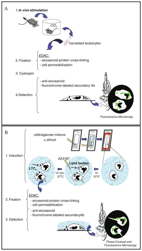
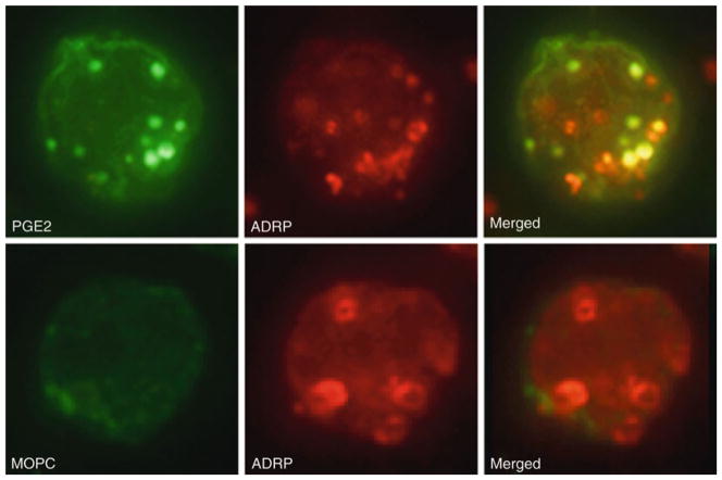
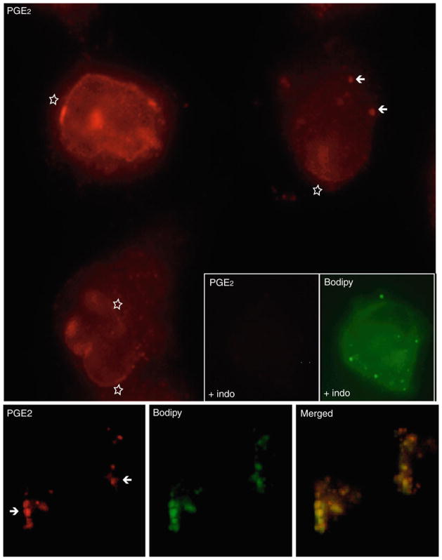
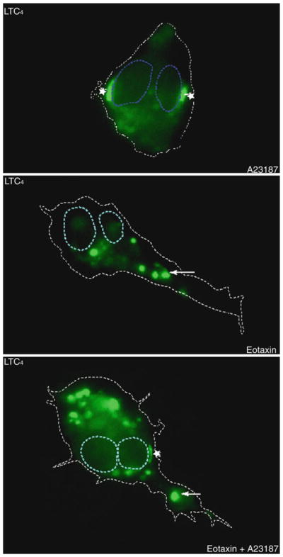
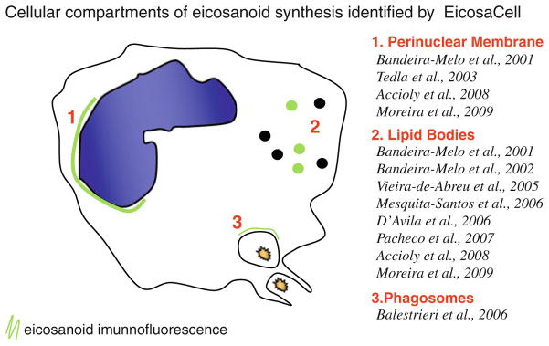
Eicosanoids (prostaglandins, leukotrienes and lipoxins) are a family of signaling lipids derived from arachidonic acid that have important roles in physiological and pathological processes. Over the past years, it has been established that successful eicosanoid production is not merely determined by arachidonic acid and eicosanoid-forming enzymes availability, but requires sequential interactions between specific biosynthetic proteins acting in cascade and may involve very unique spatial interactions. Direct assessment of specific subcellular locales of eicosanoid synthesis has been elusive, as those lipid mediators are newly formed, not stored and often rapidly released upon cell stimulation. In this chapter, we discuss the EicosaCell protocol for intracellular detection of eicosanoid-synthesizing compartments by means of a strategy to covalently cross-link and immobilize the lipid mediators at their sites of synthesis followed by immunofluorescent-based localization of the targeted eicosanoid.


Figures

Fig. 10.1
Fig. 10.1
Schematic illustration of EicosaAssay method. EicosaCell preparations, which undergo EDAC-dependent capturing and fixation of newly formed-eicosanoids at their sites of synthesis, are analyzed by phase-contrast and fluorescence microscopy and can employ cytospun cells (a), adherent cells or cells embedded in a gel matrix (b).
Fig. 10.2
Fig. 10.2
EicosaCell for PGE2 immunolocalization within BCG-infected cytospun macrophages. In upper panels, macrophages from BCG-infected animals were labeled for ADRP-associated lipid bodies (red staining) and for newly formed PGE2 (green staining). Merged image showed co-localization of PGE2 in ADRP-associated lipid bodies (yellow staining). In bottom panels, IgG1 irrelevant isotype (MOPC) was used as control for PGE2 labeling. Briefly, pleural macrophages obtained 24 h after infection with BCG were recovered from the thoracic cavity with 500 μL of HBSS, immediately mixed with 500 μL of EDAC (1% in HBSS) and incubated for 30 min at 37°C. Cells were then washed with HBSS, cytospun onto glass slides and incubated with mouse anti-PGE2 (1/100) or MOPC 21 in 0.1% normal goat serum and guinea pig polyclonal anti-mouse ADRP (1/1000) in 0.1% normal donkey serum simultaneously for 1 h at RT. Cells were washed twice and incubated with secondary Abs, goat anti-mouse conjugated with AlexaFluor-488 (1/1000) and CY3-conjugated donkey anti-guinea pig (1/1000). The slides were washed (three times, 10 min each) and mounted with aqueous mounting medium.
Fig. 10.3
Fig. 10.3
EicosaCell for PGE2 immunolocalization within adherent CACO-2 cells. The largest panel shows fluorescent microscopy of CACO-2 cells labeled for newly formed PGE2 (red staining). Bottom three images panel showed immunofluorescent PGE2 (red staining), BODIPY-associated lipid bodies (green staining) and a merged image showing co-localization of PGE2 in lipid bodies (yellow staining). Insert panel showed lack of PGE2 immunolabeling within lipid-body-enriched CACO-2 cells, which were treated with indomethacin (4 mg/kg) 1 h before EDAC. Briefly, CACO-2 cells were fixed and permeabilized during 1 h at 37°C with EDAC (0.5% in HBSS−/−). Then, cells were washed with HBSS and blocked with 2% donkey serum for 15 min before incubation with anti-PGE2 monoclonal antibody (Cayman Chemicals) for 45 min. Cells were washed with HBSS and incubated with fluorescent secondary antibody Cy3-conjugated affiniPure F(ab′) fragment donkey anti-mouse and BODIPY 493/503 (Molecular Probes, CA) for 45 min.
Fig. 10.4
Fig. 10.4
EicosaCell for LTC4 immunolabeling within gel-immersed human eosinophils. Fluorescent microscopy of agarose-embedded eosinophils, fixed with EDAC (0.5% in HBSS−/−) and stained with Alexa488-labeled anti-cysteinyl LT mAb. To facilitate intracellular localization of anti-LTC4 immunoreactive sites (green staining) within representative eosinophils, blue and white dotted lines were drawn to delineate, respectively, the nuclear and cellular perimeters. As indicated, A23187-, eotaxin- or eotaxin plus A23187-stimulated eosinophils display fluorescent LTC4 immunostaining. While, eosinophils stimulated with A23187 (0.1 μM) for 15 min exhibited exclusively perinuclear (stars) immunoreactive LTC4, eotaxin (100 ng/mL)-stimulated eosinophils showed punctate cytoplasmic lipid body-comprised LTC4 (arrows). Differently, eosinophils stimulated with eotaxin for 1 h and activated for extra 15 min with A23187 exhibit perinuclear (stars) and punctate cytoplasmic (arrows) immunoreactive LTC4.
Fig. 10.5
Fig. 10.5
Schematic summary of EiocosaCell-derived reports identifying three distinct intracellular compartments of eicosanoid synthesis: the nuclear envelope, cytoplasmic lipid bodies and zymozan-driven phagosomes.

Similar articles

Cited by

References

    1. Yaqoob P. Fatty acids as gatekeepers of immune cell regulation. Trends Immunol. 2003;24:639–645. - PubMed
    1. Wymann MP, Schneiter R. Lipid signaling in disease. Nat Rev Mol Cell Biol. 2008;9:162–176. - PubMed
    1. Smith WL, DeWitt DL, Garavito RM. Cyclooxygenases: structural, cellular, and molecular biology. Annu Rev Biochem. 2000;69:145–182. - PubMed
    1. Bozza PT, Magalhaes K, Weller PF. Leukocyte lipid bodies – biogenesis and functions in inflammation. Biochim Biophys Acta. 2009;1791:540–51. - PMC - PubMed
    1. Mandal AK, Skoch J, Bacskai BJ, Hyman BT, Christmas P, Miller D, Yamin TT, Xu S, Wisniewski D, Evans JF, Soberman RJ. The membrane organization of leukotriene synthesis. Proc Natl Acad Sci USA. 2004;101:6587–6592. - PMC - PubMed

Publication types

MeSH terms

Substances

LinkOut - more resources