. 2011 Feb;20(2):396-405.
doi: 10.1002/pro.571. Epub 2011 Jan 18.

Structures of domains I and IV from YbbR are representative of a widely distributed protein family

Affiliations

Structures of domains I and IV from YbbR are representative of a widely distributed protein family

Adam W Barb et al. Protein Sci. 2011 Feb.

Abstract

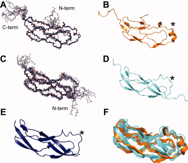
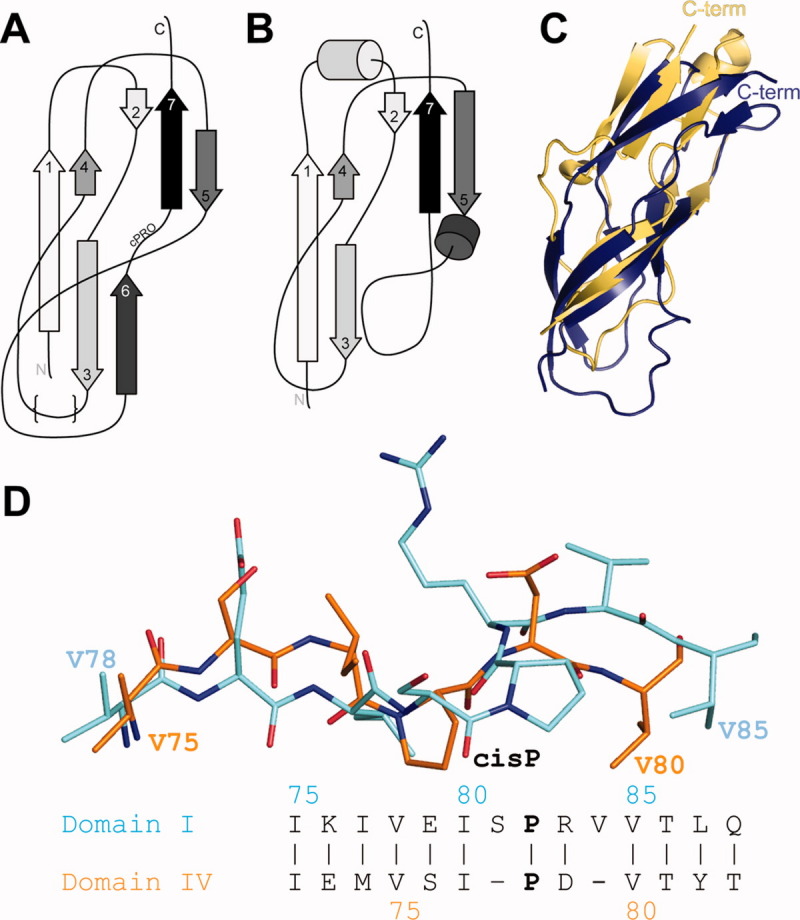
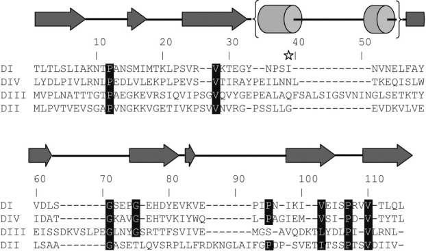
YbbR domains are widespread throughout Eubacteria and are expressed as monomeric units, linked in tandem repeats or cotranslated with other domains. Although the precise role of these domains remains undefined, the location of the multiple YbbR domain-encoding ybbR gene in the Bacillus subtilis glmM operon and its previous identification as a substrate for a surfactin-type phosphopantetheinyl transferase suggests a role in cell growth, division, and virulence. To further characterize the YbbR domains, structures of two of the four domains (I and IV) from the YbbR-like protein of Desulfitobacterium hafniense Y51 were solved by solution nuclear magnetic resonance and X-ray crystallography. The structures show the domains to have nearly identical topologies despite a low amino acid identity (23%). The topology is dominated by β-strands, roughly following a "figure 8" pattern with some strands coiling around the domain perimeter and others crossing the center. A similar topology is found in the C-terminal domain of two stress-responsive bacterial ribosomal proteins, TL5 and L25. Based on these models, a structurally guided amino acid alignment identifies features of the YbbR domains that are not evident from naïve amino acid sequence alignments. A structurally conserved cis-proline (cis-Pro) residue was identified in both domains, though the local structure in the immediate vicinities surrounding this residue differed between the two models. The conservation and location of this cis-Pro, plus anchoring Val residues, suggest this motif may be significant to protein function.


Figures

Figure. 1
Figure. 1
Structures of Domain I and IV from the Ybbr family protein of Desulfitobacterium hafniense. Panels A and C: The ensemble of 10 structures for Domain IV and I, respectively, as calculated from solution NMR spectroscopy-observed constraints. Nitrogen positions are depicted as blue sticks, and oxygen with red sticks. Panels B, D, and E: Ribbon diagrams of the lowest energy solution NMR structures of Domain IV (orange) and I (cyan) and the 1.9-Å X-ray crystal structure of Domain I (deep blue), respectively, show a unique topology that is identical for both of these domains. Panel F: An overlay of the solution structures of Domain I and IV show the similarity of these two molecules. The star marks the homology-modeled site of phosphopantetheine modification is identified by Walsh and coworkers (2005).
Figure. 2
Figure. 2
Unique structural features of Domains I and IV are conserved and are similar to the C-terminal domain of the TL5 ribosome-associated protein from Thermus thermophilus. Panel A: The unique topology of these domains forms a “figure 8”-like fold. The structurally variable region is depicted with {}s. Panel B: The topology of the TL5 protein is very similar, though one b-strand is missing and two additional helices are present. Panel C: An overlay of the Domain I X-ray structure (deep blue) and TL5 C-terminal domain (light orange) shows considerable differences. Panel D: The presence of a Pro residue preceded by a cis peptide bond is observed in structures of YbbR Domains I and IV though residues in the immediate vicinity are not conserved. However, the location of hydrophobic amino acid sidechains preceding the cis-Pro is conserved. Domain coloring is the same as in Figure 1.
Figure. 3
Figure. 3
NMR spin relaxation measurements identify dynamic regions of Domain IV that are not present in Domain I. Panel A: A measurement of the rotational correlation time of each NH moiety of Domain IV showed a relatively homogenous distribution with an average for ordered residues of 4.7 ns, suggesting ps-ns timescale motions of the backbone are uniform excepting the highly mobile C-terminal residues. Panel B: Measurements of the R2 spin relaxation rate identified three regions of Domain IV with significantly larger values, suggesting the presence slow μs-ms timescale motions. These residues likewise have greatly reduced or absent intensity in three-dimensional heteronuclear backbone assignment experiments when compared to other residues in the protein. Panel C: Similar transverse relaxation measurements of Domain I (shown) and analysis of triple-resonance experiments identified no regions with enhanced relaxation rates.
Figure. 4
Figure. 4
A structure-based sequence alignment of the four YbbR domains in the D. hafniense YbbR protein highlights regions of conservation not observed in a naïve amino acid alignment. Structurally conserved features are highlighted. Secondary structure features are shown above the alignment and are based on the domain structures presented herein. The homology modeled site of Sfp PPTase modification is shown with a star.

References

    1. Burley SK, Joachimiak A, Montelione GT, Wilson IA. Contributions to the NIH-NIGMS protein structure initiative from the PSI production centers. Structure. 2008;16:5–11. - PMC - PubMed
    1. Jaroszewski L, Li ZW, Krishna SS, Bakolitsa C, Wooley J, Deacon AM, Wilson IA, Godzik A. Exploration of uncharted regions of the protein universe. PLoS Biol. 2009:e1000205. - PMC - PubMed
    1. Finn RD, Mistry J, Tate J, Coggill P, Heger A, Pollington JE, Gavin OL, Gunasekaran P, Ceric G, Forslund K, Holm L, Sonnhammer EL, Eddy SR, Bateman A. The Pfam protein families database. Nucleic Acids Res. 2010;38:D211–D222. - PMC - PubMed
    1. Glanzmann P, Gustafson J, Komatsuzawa H, Ohta K, Berger-Bachi B. glmM operon and methicillin-resistant glmM suppressor mutants in Staphylococcus aureus. Antimicrob Agents Chemother. 1999;43:240–245. - PMC - PubMed
    1. Mengin-Lecreulx D, vanHeijenoort J. Characterization of the essential gene glmM encoding phosphoglucosamine mutase in Escherichia coli. J Biol Chem. 1996;271:32–39. - PubMed

Publication types

Substances

LinkOut - more resources