Review
. 2011 Jan 1;71(1):45-61.
doi: 10.1002/dneu.20853.

Three groups of interneurons account for nearly 100% of neocortical GABAergic neurons

Affiliations
Review

Three groups of interneurons account for nearly 100% of neocortical GABAergic neurons

Bernardo Rudy et al. Dev Neurobiol. .

Abstract

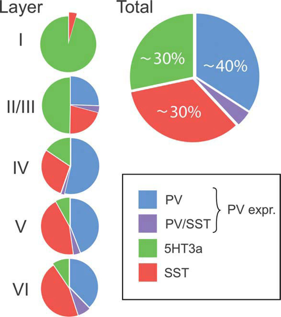
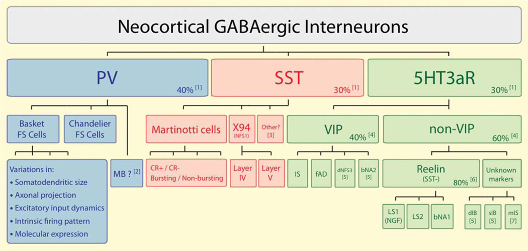
An understanding of the diversity of cortical GABAergic interneurons is critical to understand the function of the cerebral cortex. Recent data suggest that neurons expressing three markers, the Ca2+-binding protein parvalbumin (PV), the neuropeptide somatostatin (SST), and the ionotropic serotonin receptor 5HT3a (5HT3aR) account for nearly 100% of neocortical interneurons. Interneurons expressing each of these markers have a different embryological origin. Each group includes several types of interneurons that differ in morphological and electrophysiological properties and likely have different functions in the cortical circuit. The PV group accounts for ∼40% of GABAergic neurons and includes fast spiking basket cells and chandelier cells. The SST group, which represents ∼30% of GABAergic neurons, includes the Martinotti cells and a set of neurons that specifically target layerIV. The 5HT3aR group, which also accounts for ∼30% of the total interneuronal population, is heterogeneous and includes all of the neurons that express the neuropeptide VIP, as well as an equally numerous subgroup of neurons that do not express VIP and includes neurogliaform cells. The universal modulation of these neurons by serotonin and acetylcholine via ionotropic receptors suggests that they might be involved in shaping cortical circuits during specific brain states and behavioral contexts.


Figures

Figure 1
Figure 1
Distribution of three groups of GABAergic interneurons in S1 cortex. Shown are the proportions of GAD-67 mRNA-expressing neurons also expressing mRNA for parvalbumin (PV), somatostatin (SST), or 5HT3aR based on the study by Lee et al. (2010). PV-expressing interneurons (including those expressing SST mRNA) account for ~40% of the total GABAergic population in the mouse S1 cortex. SST-expressing and 5HT3aR-expressing neurons account for ~30% each. The distribution of interneuron groups varies among different cortical layers. 5HT3aR neurons are concentrated in supragranular layers. Layer I, which has a much lower cell density, expresses mainly 5HT3aR neurons. In layer I and in layers II/III, the proportion of 5HT3aR neurons exceeds the proportion of PV cells. The proportion of PV cells is particularly large in layer IV. There are no PV neurons in layer I. SST interneurons are found in all layers, but are particularly abundant in infragranular layers.
Figure 2
Figure 2
A classification of neocortical GABAergic interneurons. Nearly 100% of all neocortical GABAergic neurons belong to one of three groups defined by the expression of parvalbumin (PV), somatostatin (SST), and the ionotropic serotonin receptor 5HT3a (5HT3aR). Each group consists of several subgroups. In turn, each subgroup consists of several functionally distinct types or classes of interneurons, most of which are still poorly defined. PV expression has been associated with the fast-spiking (FS) firing pattern. There are two anatomically distinct subgroups of FS neurons, basket cells, and chandelier cells. MB (multipolar bursting) neurons define a possible subgroup of non-FS, PV-expressing neurons. FS basket cells have been observed to vary in a number of properties, but it is not clear which or how many classes of FS basket cells are there in neocortex. Two subgroups of SST neurons are well characterized, Martinotti cells, which account for the majority of SST neurons, and a subgroup of neurons described in the X94 mouse line characterized by a profuse axonal projection to layer IV. Shown as “other” are SST neurons that do not seem to belong to any of these two categories (McGarry et al., 2010). Martinotti cells have been shown to vary in a number of morphological, electrophysiological, and molecular properties, but it is not possible yet to clearly define classes of Martinotti cells. For example, Martinotti cells can be subdivided into those that express calretinin (CR+) and those that do not (CR−), which vary in a number of properties (see text). X94 cells are present in layer IV and V, both of which project to layer IV. The subdivisions of the 5HT3aR group are still poorly defined. 5HT3aR neurons can be classified into two subgroups based on the expression of the neuropeptide VIP. Each of these subgroups of 5HT3aR neurons consists of several, still mostly undefined, types of interneurons. The most frequently observed types include, among the VIP neurons, the bipolar or bitufted neurons with an irregular spiking (IS) or a “fast adapting” (fAD) firing pattern, and among the non-VIP neurons, the neurogliaform (NGF) cells with a late spiking firing pattern (LS1) and multipolar neurons with an LS2 firing pattern. Note: (1) From Lee et al. (2010); (2) See Blatow et al. (2003); (3) See text for SST neurons described by McGarry et al. (2010); (4) Estimates based on Miyoshi et al. (2010) and Lee et al. (2010); (5) Neurons with a dFNS3, bNA2, dIB, and sIB firing patterns were more rarely sampled than other CGE-derived neurons by Miyoshi et al. (2010); (6) Reelin, which is also expressed by a fraction of SST neurons, was shown to be expressed in a major fraction of non-VIP CGE-derived (Miyoshi et al., 2010) and 5HT3aR (Lee et al., 2010) neurons; (7) A fraction of non-VIP 5HT3aR neurons (mIS) had multipolar morphology and an IS firing pattern (Lee et al., 2010) resembling neurons derived from the POA (see text).

References

    1. Acsady L, Gorcs TJ, Freund TF. Different populations of vasoactive intestinal polypeptide-immunoreactive interneurons are specialized to control pyramidal cells or interneurons in the hippocampus. Neuroscience. 1996;73:317–334. - PubMed
    1. Anderson SA, Eisenstat DD, Shi L, Rubenstein JL. Interneuron migration from basal forebrain to neocortex: Dependence on Dlx genes. Science. 1997;278:474–476. - PubMed
    1. Ascoli GA, Alonso-Nanclares L, Anderson SA, Barrio-nuevo G, Benavides-Piccione R, Burkhalter A, Buzsaki G, et al. Petilla terminology: Nomenclature of features of GABAergic interneurons of the cerebral cortex. Nat rev. 2008;9:557–568. - PMC - PubMed
    1. Bacci A, Huguenard JR, Prince DA. Modulation of neocortical interneurons: Extrinsic influences and exercises in self-control. Trends Neurosci. 2005;28:602–610. - PubMed
    1. Bartos M, Vida I, Jonas P. Synaptic mechanisms of synchronized gamma oscillations in inhibitory interneuron networks. Nat Rev. 2007;8:45–56. - PubMed

Substances