. 2011 Feb 1;50(4):502-11.
doi: 10.1021/bi101059w. Epub 2010 Dec 30.

Direct measurement of thermal stability of expressed CCR5 and stabilization by small molecule ligands

Affiliations

Direct measurement of thermal stability of expressed CCR5 and stabilization by small molecule ligands

Adam M Knepp et al. Biochemistry. .

Abstract

The inherent instability of heptahelical G protein-coupled receptors (GPCRs) during purification and reconstitution is a primary impediment to biophysical studies and to obtaining high-resolution crystal structures. New approaches to stabilizing receptors during purification and screening reconstitution procedures are needed. Here we report the development of a novel homogeneous time-resolved fluorescence assay (HTRF) to quantify properly folded CC-chemokine receptor 5 (CCR5). The assay permits high-throughput thermal stability measurements of femtomole quantities of CCR5 in detergent and in engineered nanoscale apolipoprotein-bound bilayer (NABB) particles. We show that recombinantly expressed CCR5 can be incorporated into NABB particles in high yield, resulting in greater thermal stability compared with that of CCR5 in a detergent solution. We also demonstrate that binding of CCR5 to the HIV-1 cellular entry inhibitors maraviroc, AD101, CMPD 167, and vicriviroc dramatically increases receptor stability. The HTRF assay technology reported here is applicable to other membrane proteins and could greatly facilitate structural studies of GPCRs.


Figures

FIGURE 1
FIGURE 1
(A) Scheme depicting strategy to synthesize EuK-labeled 2D7 mAb (9). EuK (1) is activated by addition of a sulfhydryl by reaction with SPDP (2) and reduction by TCEP (top). Separately, 2D7 mAb (6) is immobilized on Ni-NTA resin and reacted with sulfo-SMCC (7) to generate a maleimide derivative (8). A crystal structure of IgG (PDB ID: 1IGY) is shown as a model. The two activated reagents are combined and labeled 2D7 is eluted from the resin with imidazole. (B) Left: Size-exclusion chromatography with monitoring of absorbance at 280 nm (blue; protein) and 305 nm (red; EuK) to determine yield and labeling ratio. Right: Coomassie blue-stained non-reducing SDS polyacrylamide gel electrophoresis showing a single band of the intact 2D7-EuK after labeling. Note the impurities of the initial 2D7 sample have been largely removed by the procedure.
FIGURE 2
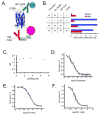
FIGURE 2
(A) HTRF sandwich immunoassay schematic. A hypothetical model of CCR5 based on the crystal structure of rhodopsin (PDB ID: 1U19) is shown. 2D7-EuK recognizes a conformation-sensitive split epitope on the extracellular side of CCR5. Biotinylated 1D4 (1D4-biot) binds an engineered nine-residue C-terminal epitope (red) and is linked to streptavidin-conjugated XL665. FRET is observed between EuK and XL665 when 2D7 binds properly folded CCR5. The Förster radius for this donor-acceptor pair is approximately 95 Å. (B) Assay controls with fluorescence counts at 615 nm (blue) and 665 nm (red) after excitation at 320 nm. CCR5-specific signal is seen as a signal increase at 665 nm and decrease at 615 nm. (C) A serial dilution of CCR5 shows the dynamic range of the assay. The signal saturates at ∼200% enhancement over background. ΔF is defined as: ΔF=(F665,sampleF615,sampleF665,negativeF615,negative)÷F665,negativeF615,negative (D-F) Competition experiments with the 1D5 nonapeptide (D), 1D4 mAb (E), and 2D7 mAb (F) demonstrate signal specificity. The apparent IC50 values are 130 nM, 2.7 nM, and 0.79 nM for the three competitors, respectively.
FIGURE 3
FIGURE 3
(A) Chromatogram showing co-elution of protein (blue; 280 nm) and fluorescent lipids (red; 570 nm). The 1D4 immunoblot below shows that CCR5-NABBs elute in the peak centered at 15.6 mL, exhibiting larger hydrodynamic radius than the majority of the NABBs at 16.1 and 17.0 mL. The anti-His6 blot detects His-tagged zap1 belt protein. Free zap1 elutes at 18.6 mL (inset). (B) CCR5-NABBs were incubated with Protein G beads and 2D7, and the supernatant fraction was probed with a 1D4 immunoblot. The majority of CCR5 in NABBs is immunoprecipitated by 2D7, indicating properly folded receptor. (C) HTRF signal from a serial dilution of CCR5-NABBs, demonstrating the ability to quantify properly folded CCR5 in NABBs. (D) The HTRF signal is efficiently competed with 1 μM 1D4, showing signal specificity.
FIGURE 4
FIGURE 4
The HTRF assay was applied to make high-throughput thermal stability measurements with femtomole quantities of CCR5. A range of temperatures was applied to detergent-solubilized CCR5 or CCR5-NABBs before adding to labeled HTRF components in a 384-well plate. (A) Melting curves of unliganded CCR5 (black) and CCR5 preincubated with the small molecule antagonist maraviroc (red). Maraviroc shifts the TM of detergent-solubilized CCR5 from 47.1°C to 66.0°C (rightward-pointing arrow) and appears to display a two-step binding profile. The first ligand-receptor state reduces the accessibility of the 2D7 epitope on the EC2 loop compared with unliganded receptor (downward-pointing arrow). The second ligand-receptor state results in a higher HTRF signal (upward-pointing arrow). (B) Thermal denaturation of CCR5-ligand complexes. CCR5 was preincubated with maraviroc (red), AD101 (green), vicriviroc (blue), and CMPD 167 (cyan). All of these antagonists appear to have two binding states. The calculated TM for maraviroc, AD101, vicriviroc, and CMPD 167 is 66.0°C, 59.9°C, 59.5°C, and 62.7°C, respectively. The unliganded receptor, which melts at a lower temperature, is shown as a black dashed line for reference. (C) Molecular structures of the CCR5 antagonists tested. (D) Melting curve of CCR5-NABBs. The assembly denatures at 54.5°C. CCR5-NABBs melt over a much broader range than CCR5 in detergent solution, suggesting some sample heterogeneity or a distinct denaturation pathway.

References

    1. Deupi X, Kobilka B. Mechanisms and Pathways of Heterotrimeric G Protein Signaling. Elsevier Academic Press Inc; San Diego: 2007. Activation of G Protein Coupled Receptors; pp. 137–166.
    1. Premont RT, Gainetdinov RR. Physiological roles of G protein-coupled receptor kinases and arrestins. Annu Rev Physiol. 2007;69:511–534. - PubMed
    1. Lagerstrom MC, Schioth HB. Structural diversity of G protein-coupled receptors and significance for drug discovery. Nat Rev Drug Discov. 2008;7:339–357. - PubMed
    1. Rosenbaum DM, Rasmussen SGF, Kobilka BK. The structure and function of G-protein-coupled receptors. Nature. 2009;459:356–363. - PMC - PubMed
    1. Bartfai T, Benovic JL, Bockaert J, Bond RA, Bouvier M, Christopoulos A, Civelli O, Devi LA, George SR, Inui A, Kobilka B, Leurs R, Neubig R, Pin JP, Quirion R, Roques BP, Sakmar TP, Seifert R, Stenkamp RE, Strange PG. The state of GPCR research in 2004. Nat Rev Drug Discov. 2004;3:574–626. - PubMed

Publication types

MeSH terms