. 2011 Jan 25;5(1):237-46.
doi: 10.1021/nn1024658. Epub 2010 Dec 14.

Fabrication of stable and RNase-resistant RNA nanoparticles active in gearing the nanomotors for viral DNA packaging

Affiliations

Fabrication of stable and RNase-resistant RNA nanoparticles active in gearing the nanomotors for viral DNA packaging

Jing Liu et al. ACS Nano. .

Abstract

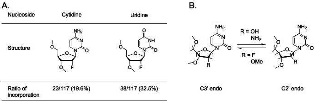
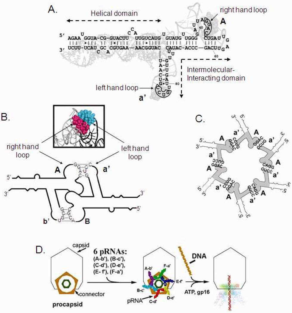
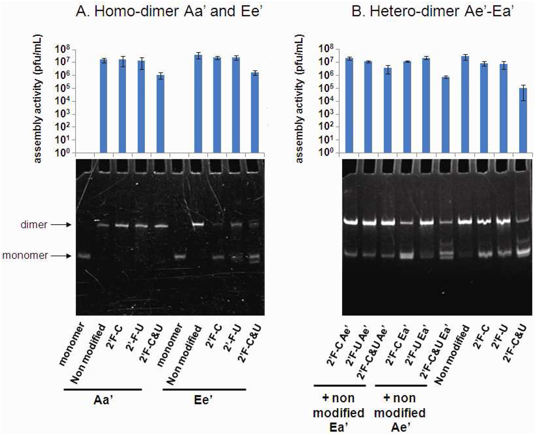
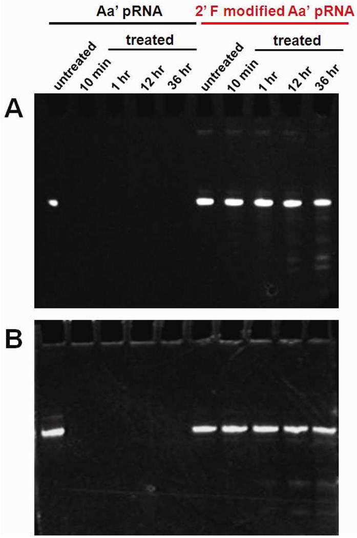
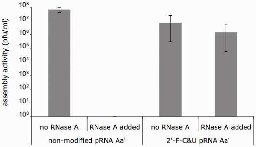
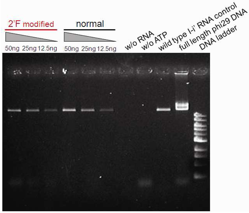
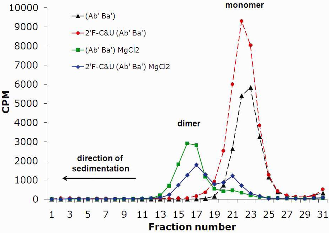
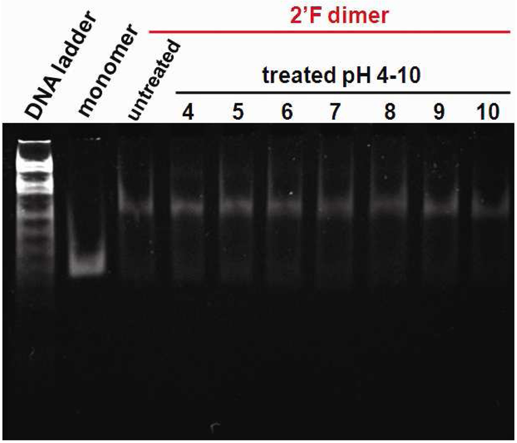
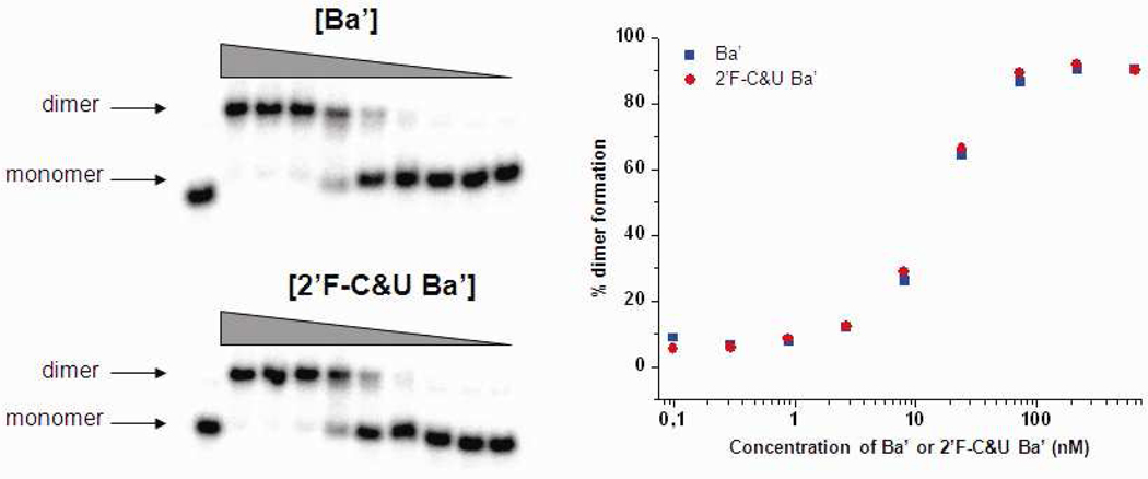
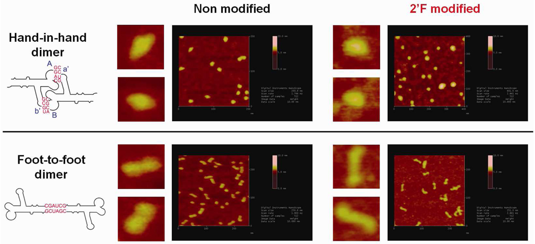
Both DNA and RNA can serve as powerful building blocks for bottom-up fabrication of nanostructures. A pioneering concept proposed by Ned Seeman 30 years ago has led to an explosion of knowledge in DNA nanotechnology. RNA can be manipulated with simplicity characteristic of DNA, while possessing noncanonical base-pairing, versatile function, and catalytic activity similar to proteins. However, standing in awe of the sensitivity of RNA to RNase degradation has made many scientists flinch away from RNA nanotechnology. Here we report the construction of stable RNA nanoparticles resistant to RNase digestion. The 2'-F (2'-fluoro) RNA retained its property for correct folding in dimer formation, appropriate structure in procapsid binding, and biological activity in gearing the phi29 nanomotor to package viral DNA and producing infectious viral particles. Our results demonstrate that it is practical to produce RNase-resistant, biologically active, and stable RNA for application in nanotechnology.


Figures

Figure 1
Figure 1
Structure of the 2’-modified ribonucleotides. (A) Location of modification and ratio of incorporation in the pRNA Aa’ sequence. (B) Illustration of the equilibrium between the C2’ endo and C3’ endo conformation of the sugar ring following the 2’ substituent.
Figure 2
Figure 2
Sequence and secondary structural of phi29 DNA-packaging RNA (pRNA). Upper-case letters represent the upper loop and lower-case letters the lower loop of pRNA. A pair of upper and lower case for a same letters indicate a pair of complementary loops, whereas a pair of upper and lower case from different letters indicate non-complementary loops. For example, pRNA Aa’ refers to a pRNA with complementary right loop A (5’G45G46A47C48) and left loop a’ (3’C85C84U83G82), which can form homo-dimer by itself. pRNA Ae’ containing non-complementary upper loop A (5’G45G46A47C48) and lower loop e’ (3’C85G84G83U82) can form hetero-dimer with pRNA Ea’ containing left-hand loop a’ (3’C85C84U83G82) and right-hand loop E (5’G45C46C47A48). (A) Superposition of the primary and secondary structure of the pRNA Aa’. The four bases in the right and left loops, which are responsible for inter-RNA interactions are boxed. (B) Diagram depicting pRNA dimer formation and 3D model of the hand-in-hand interaction. (C) pRNA hexamer. (D) Packaging of phi29 DNA through the motor geared by six pRNA.
Figure 3
Figure 3
Native PAGE gel showing the formation of dimer nanoparticle of pRNA and histogram of the virion assembly activities associated to these nanoparticle (A) homodimer Aa’ and Ee’. (B) Hetero-dimer Ae’/Ea’.
Figure 4
Figure 4
Urea-PAGE denatured gel showing the stability between non-modified pRNA Aa’ and 2’-F-C&U pRNA Aa’ after incubation at different time point in the presence of (A) RNase A (1 mg/ml), and (B) fetal bovine serum (10%).
Figure 5
Figure 5
Assembly activities of the 2’-F-C&U pRNA Aa’ and its non-modifed counterparts in the presence or absence of RNase A.
Figure 6
Figure 6
Agarose gel showing the efficiency of the 2’-F-C&U pRNA Aa’ in DNA packaging compared to its non-modified counterparts.
Figure 7
Figure 7
Sucrose gradient sedimentation analysis of the formation of [3H]pRNA dimer in presence or absence of MgCl2.
Figure 8
Figure 8
Native PAGE gel showing the stability of the 2’-F-C&U pRNA Aa’ homo-dimer after incubation at different pH ranging from 4 to 10.
Figure 9
Figure 9
Titration of dimer formation between [32P] prNA Ab’ and cold 2’-F-C&U and non-modified pRNA Ba’ (0.1, 0.3, 0.9, 2.6, 8, 24, 72, 216 and 650 nM). (A) Native PAGE gel. (B) Plot showing the percentage of dimer formation function of the concentration of cold 2’-F-C&U and non-modified pRNA Ba’.
Figure 10
Figure 10
Atomic force microscopy images showing hand-in-hand and foot-to-foot dimer nanoparticle of non-modified pRNA and 2’-F-C&U pRNA respectively.

Similar articles

Cited by

References

    1. Lin C, Liu Y, Yan H. Designer DNA Nanoarchitectures. Biochemistry. 2009;48:1663–1674. - PMC - PubMed
    1. Aldaye FA, Palmer AL, Sleiman HF. Assembling Materials With DNA As the Guide. Science. 2008;321:1795–1799. - PubMed
    1. Seeman NC. Nanomaterials Based on DNA. Annu. Rev. Biochem. 2010;79:65–87. - PMC - PubMed
    1. Glotzer SC. Materials Science. Some Assembly Required. Science. 2004;306:419–420. - PubMed
    1. Gates BD, Xu Q, Stewart M, Ryan D, Willson CG, Whitesides GM. New Approaches to Nanofabrication: Molding, Printing, and Other Techniques. Chem. Rev. 2005;105:1171–1196. - PubMed

Publication types

MeSH terms