Review
. 2011 May;15(5):1013-31.
doi: 10.1111/j.1582-4934.2010.01236.x.

Proteoglycans in cancer biology, tumour microenvironment and angiogenesis

Affiliations
Review

Proteoglycans in cancer biology, tumour microenvironment and angiogenesis

Renato V Iozzo et al. J Cell Mol Med. 2011 May.

Abstract

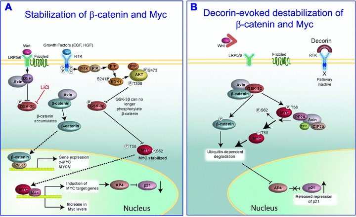
Proteoglycans, key molecular effectors of cell surface and pericellular microenvironments, perform multiple functions in cancer and angiogenesis by virtue of their polyhedric nature and their ability to interact with both ligands and receptors that regulate neoplastic growth and neovascularization. Some proteoglycans such as perlecan, have pro- and anti-angiogenic activities, whereas other proteoglycans, such as syndecans and glypicans, can also directly affect cancer growth by modulating key signalling pathways. The bioactivity of these proteoglycans is further modulated by several classes of enzymes within the tumour microenvironment: (i) sheddases that cleave transmembrane or cell-associated syndecans and glypicans, (ii) various proteinases that cleave the protein core of pericellular proteoglycans and (iii) heparanases and endosulfatases which modify the structure and bioactivity of various heparan sulphate proteoglycans and their bound growth factors. In contrast, some of the small leucine-rich proteoglycans, such as decorin and lumican, act as tumour repressors by physically antagonizing receptor tyrosine kinases including the epidermal growth factor and the Met receptors or integrin receptors thereby evoking anti-survival and pro-apoptotic pathways. In this review we will critically assess the expanding repertoire of molecular interactions attributed to various proteoglycans and will discuss novel proteoglycan functions modulating cancer progression, invasion and metastasis and how these factors regulate the tumour microenvironment.


Figures

fig 1
fig 1
Schematic representation of the dual activity of pelican. Perlecan can act as a powerful pro-angiogenic factor by either directly presenting VEGFA (and other heparan sulphate growth factors) to the VEGFR2, or indirectly following partial heparanase cleavage of the heparan sulphate side chains. Both can trigger VEGFR2 signalling with stimulation of migration, vascular permeability, survival and proliferation. Perlecan can also act as a powerful anti-angiogenic factor following cathepsin L or BMP-1/Tolloid-like protease cleavage of endorepellin and LG3, respectively. These C-terminal fragments bind with high affinity to the α2 I domain of the α2β1 integrin triggering a signal cascade that leads to disruption of endothelial cytoskeleton and endothelial cell motility. Endorepellin also activates the tyrosine phosphatase SHP-1 which is bound to the cytoplasmic domain of the α2β1 integrin. SHP-1 then dephosphorylates a number of RTKs including VEGFR2, thereby blocking endothelial cell migration, survival and proliferation.
fig 2
fig 2
Heparanase enhances gene expression and syndecan-1 (SDC1) shedding which together enhance tumour progression. Upon up-regulation of heparanase expression by the tumour cell, expression of syndecan-1, MMP-9, VEGF, HGF and receptor activator of nuclear factor κ-B ligand (RANKL) are elevated and released into the tumour microenvironment. MMP-9 acts as a ‘sheddase’ to increase release of syndecan-1 from the tumour cell surface (1). Shed syndecan-1 binds to VEGF (2) to promote angiogenesis and to HGF (3) to stimulate tumour growth. RANKL (4) activates differentiation of osteoclast precursors to accelerate bone turnover leading to release of factors that further drive tumour progression. Heparanase also cleaves the heparan sulphate chains of perlecan (5) thereby liberating additional VEGF that further stimulates angiogenesis.
fig 3
fig 3
Schematic representation of decorin’s effects on β-catenin and Myc stabilization by directly down-regulating EGFR and Met. (A) Activation of both the Wnt and RTK pathways leads to stabilization of β-catenin and Myc proteins and their subsequent translocation into the nuclei where downstream transcription is activated. (B) When the Wnt pathway is blocked by absence or block of Wnt factors, or when RTK activity is attenuated by exogenous decorin, there is phosphorylation of β-catenin and Myc at specific residues. Especially important is phosphorylation at Threonine 58 (T58) of Myc, because this modification acts as priming for subsequent ubiquitination and 26S proteasomal degradation. For additional details see the text. Adapted in part from Albihn et al.

References

    1. Adany R, Heimer R, Caterson B, et al. Altered expression of chondroitin sulfate proteoglycan in the stroma of human colon carcinoma. Hypomethylation of PG-40 gene correlates with increased PG-40 content and mRNA levels. J Biol Chem. 1990;265:11389–96. - PubMed
    1. Iozzo RV. Tumor stroma as a regulator of neoplastic behavior. Agonistic and antagonistic elements embedded in the same connective tissue. Lab Invest. 1995;73:157–60. - PubMed
    1. Iozzo RV. Heparan sulfate proteoglycans: intricate molecules with intriguing functions. J Clin Invest. 2001;108:165–7. - PMC - PubMed
    1. Iozzo RV. Basement membrane proteoglycans: from cellar to ceiling. Nature Rev Mol Cell Biol. 2005;6:646–56. - PubMed
    1. Iozzo RV, Zoeller JJ, Nyström A. Basement membrane proteoglycans: modulators par excellence of cancer growth and angiogenesis. Mol Cells. 2009;27:503–13. - PMC - PubMed

Publication types