. 2010 Dec 15;99(12):3941-50.
doi: 10.1016/j.bpj.2010.10.024.

Local geometry and elasticity in compact chromatin structure

Affiliations

Local geometry and elasticity in compact chromatin structure

Elena F Koslover et al. Biophys J. .

Abstract

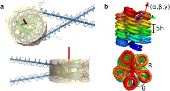
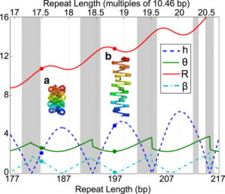
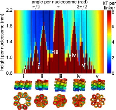
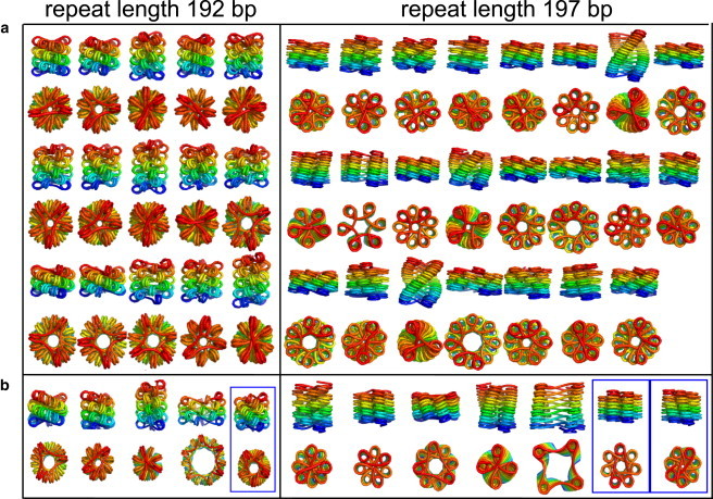
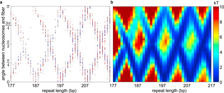
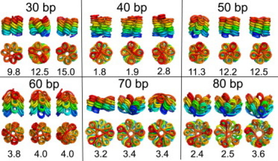
The hierarchical packaging of DNA into chromatin within a eukaryotic nucleus plays a pivotal role in both the accessibility of genomic information and the dynamics of replication. Our work addresses the role of nanoscale physical and geometric properties in determining the structure of chromatin at the mesoscale level. We study the packaging of DNA in chromatin fibers by optimization of regular helical morphologies, considering the elasticity of the linker DNA as well as steric packing of the nucleosomes and linkers. Our model predicts a broad range of preferred helix structures for a fixed linker length of DNA; changing the linker length alters the predicted ensemble. Specifically, we find that the twist registry of the nucleosomes, as set by the internucleosome repeat length, determines the preferred angle between the nucleosomes and the fiber axis. For moderate to long linker lengths, we find a number of energetically comparable configurations with different nucleosome-nucleosome interaction patterns, indicating a potential role for kinetic trapping in chromatin fiber formation. Our results highlight the key role played by DNA elasticity and local geometry in regulating the hierarchical packaging of the genome.


Figures

Figure 1
Figure 1
(a) Nucleosome geometry as obtained from crystallographic structure. (Beige cylinder) Shape of the nucleosome used for steric exclusion. (Red) Symmetry axis for the nucleosome. (b) The six coordinates used to specify a regular helix fiber structure. Because this is a five-stack structure, the height between each nucleosome and the one above it corresponds to 5h.
Figure 2
Figure 2
Superhelical parameters for straight-linker structures with different linker lengths. (Blue dashed line) Height per nucleosome (h) in nm. (Green solid line) Angle per nucleosome (θ) in radians. (Red solid line) Radius (R) in nm. (Cyan dash-dotted line) Angle of nucleosome relative to fiber axis (β) in radians. (Gray regions) Sterically excluded. Example structures are shown for (a) 36-bp and (b) 50-bp linker lengths.
Figure 3
Figure 3
Profile of minimal energies for fiber structures with 50-bp linkers, as a function of height and angle per nucleosome. (Vertical lines) Angles that are small rational multiples of 2π and thus lead to steric clashes between nucleosomes. (Bottom) Example structures.
Figure 4
Figure 4
(a) Side and top views of all candidate structures for compact fibers with crystallographic nucleosome geometry, 192-bp and 197-bp repeat lengths (45-bp and 50-bp linkers, respectively). The structures shown are local minima in all coordinates other than the height per nucleosome. Only structures with height per nucleosome below 2.2 nm and energy below 3.4 kT are shown. Parameters are listed in Table S1 in the Supporting Material. (b) Example candidate structures for nucleosomes with 10 bp unwrapped at each end. Boxed structures have topologically entwined linkers.
Figure 5
Figure 5
(a) Angle (β) between nucleosomes and fiber axis for all candidate structures at all repeat lengths. (Blue) Crystallographic geometry. (Red) Ten basepairs unwrapped at each edge. (b) Profile of minimal energies to form a structure with the given β-angle for different repeat lengths.
Figure 6
Figure 6
Top three structures for fibers of different linker lengths (30–90 bp) with height per nucleosome and fiber radius constrained to values measured by electron microscopy (4). The energy for each locally optimized structure is given in kT per linker.

References

    1. Richmond T.J., Davey C.A. The structure of DNA in the nucleosome core. Nature. 2003;423:145–150. - PubMed
    1. Huynh V.A., Robinson P.J., Rhodes D. A method for the in vitro reconstitution of a defined “30 nm” chromatin fiber containing stoichiometric amounts of the linker histone. J. Mol. Biol. 2005;345:957–968. - PubMed
    1. Langmore J.P., Schutt C. The higher order structure of chicken erythrocyte chromosomes in vivo. Nature. 1980;288:620–622. - PubMed
    1. Robinson P.J.J., Fairall L., Rhodes D. EM measurements define the dimensions of the “30-nm” chromatin fiber: evidence for a compact, interdigitated structure. Proc. Natl. Acad. Sci. USA. 2006;103:6506–6511. - PMC - PubMed
    1. Kruithof M., Chien F.T., van Noort J. Single-molecule force spectroscopy reveals a highly compliant helical folding for the 30-nm chromatin fiber. Nat. Struct. Mol. Biol. 2009;16:534–540. - PubMed

Publication types

LinkOut - more resources