. 2011 Aug;55(2):399-406.
doi: 10.1016/j.jhep.2010.11.025. Epub 2010 Dec 13.

Ethanol-stimulated differentiated functions of human or mouse hepatic stellate cells are mediated by connective tissue growth factor

Affiliations

Ethanol-stimulated differentiated functions of human or mouse hepatic stellate cells are mediated by connective tissue growth factor

Li Chen et al. J Hepatol. 2011 Aug.

Abstract

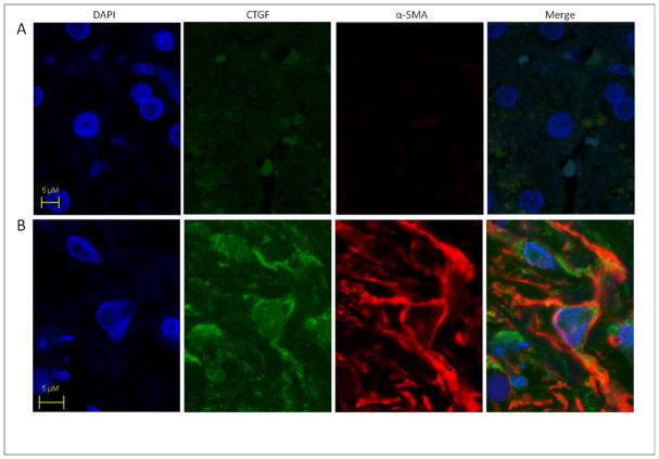
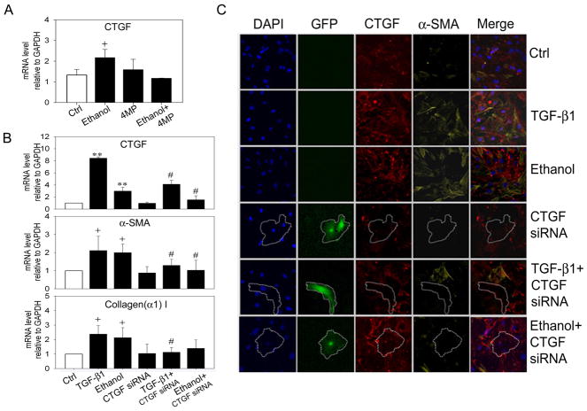
Background & aims: Connective tissue growth factor (CTGF) expression is intimately associated with hepatic fibrotic pathophysiology. In this study, CTGF production and action was investigated in ethanol-treated mouse primary hepatic stellate cells (HSC) or human LX-2 cells.

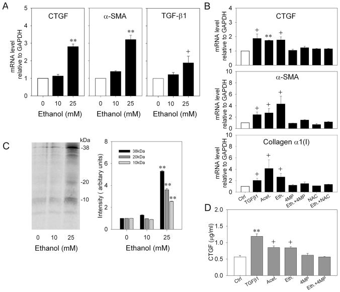
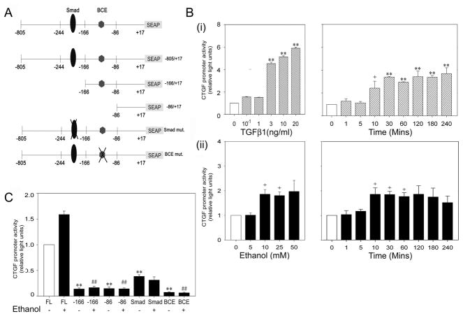
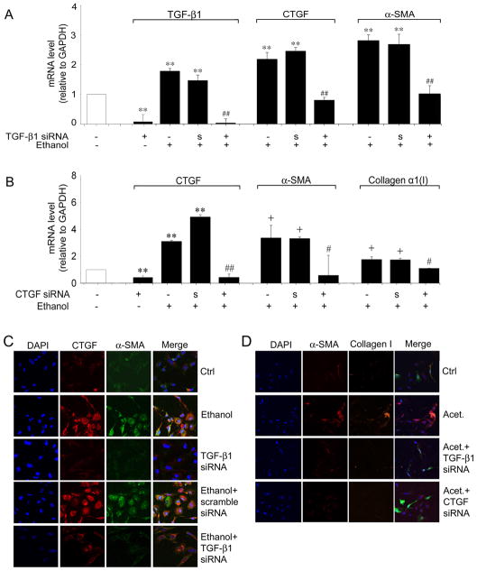
Methods: CTGF, transforming growth factor-beta1 (TGF-β1), alpha-smooth muscle actin (α-SMA) or collagen α1(I) mRNA were quantified by real-time PCR after treatment of HSC with ethanol or acetaldehyde. CTGF protein production was assessed by immunoprecipitation or ELISA. Ethanol-stimulated CTGF transcription was investigated using CTGF promoter reporter constructs. The TGF-β1- or CTGF-dependency of ethanol-induced CTGF, α-SMA, or collagen α1(I) was determined using small interfering RNA (siRNA) to TGF-β1 or CTGF.

Results: In human steatohepatitis, CTGF was produced by presumptive activated HSC. In cultured human or mouse HSC, production of CTGF, α-SMA and/or collagen was increased by ethanol treatment, an effect mimicked by acetaldehyde and blocked by 4-methylpyrazole (4-MP) or N-acetylcysteine (NAC). CTGF promoter activity was stimulated in a sustained fashion by ethanol or TGF-β1. Mutation of the Smad site or basal control element (BCE-1) in the CTGF promoter caused a 5-fold reduction in ethanol-stimulated CTGF promoter activity. Administration of TGF-β1 siRNA or CTGF siRNA significantly decreased ethanol- or acetaldehyde-stimulated mRNA or protein levels of CTGF, α-SMA or collagen I in LX-2 cells. In mouse HSC, TGF-β1- or ethanol-stimulated CTGF, α-SMA or collagen I were significantly attenuated by CTGF siRNA.

Conclusions: Ethanol-induced α-SMA or collagen α1(I) in HSC are mediated via TGF-β-dependent CTGF production, highlighting potential therapeutic benefits of targeting CTGF in alcoholic liver disease.


Figures

Fig. 1
Fig. 1. CTGF production by myofibroblasts in human steatohepatitis
Sections of (A) normal human liver or (B) steatohepatitis with moderate activity and septal fibrosis stage 3 were stained with DAPI for nuclear localization and processed for CTGF and α-SMA immunohistochemistry. Images were merged to verify co-localization of CTGF and α-SMA to the same myofibroblastic cells (i.e. presumptive activated HSC) within the fibrotic regions (yellow-orange color). Data are representative of six samples of human alcoholic liver fibrosis.
Fig.2
Fig.2. Effect of ethanol, TGF-β1 or acetaldehyde on CTGF, TGF-β1, α-SMA or collagen α 1(I) expression in LX-2 cells
(A.B) Cultured LX-2 cells were serum-starved for 24h prior to 48-hour treatment with 0–25mM ethanol ± 2mM 4MP ± 1mM NAC, 5ng/ml TGF-β1 or 100μM acetaldehyde. RNA was subjected to real-time PCR and expression of CTGF, TGF-β1, α-SMA or collagen α 1(I) was normalized to that of GAPDH. (**p<0.001, + p<0.05 v no treatment). (C) LX-2 cells were labeled with 100μCi /ml [35S] methionine/cysteine in the presence or absence of 0–25mM ethanol for 48h. CTGF immunoprecipitates were run on 18% SDS-PAGE gels and analyzed by autoradiography. The figure shows a representative blot (left) and quantification of three independent experiments (right). (**p<0.001 v no ethanol). (D) Lysates from serum-starved LX-2 cells treated for 48h with 0–25mM ethanol ± 2mM 4MP ± 1mM NAC, 5ng/ml TGF-β1 or 100μM acetaldehyde were assayed for CTGF content by ELISA. (**p<0.001, + p<0.05 v Ctrl).
Fig.3
Fig.3. Transcriptional response elements in the CTGF promoter that drive ethanol-stimulated reporter activity in LX-2 cells
(A) Organization of CTGF promoter (−805 to +17), fused to a SEAP reporter gene showing the location of key transcriptional elements that were altered by point mutation or deletion mutation. (B) Cultured LX-2 cells were transfected with plasmids containing the CTGF promoter-reporter, serum-starved for 18h, and then treated with (i) 0–20 ng/ml TGF-β1 for 6h (left) or 10 ng/ml TGF-β1 for 0–240 mins (right) or (ii) 0–50 mM ethanol for 2h (left) or 25 mM ethanol for 0–240 mins (right). SEAP reporter activity was calculated after adjustment for differences among samples in transfection efficiency as determined by co-transfection with a β-gal reporter. (**p<0.001, +p<0.05 v no treatment). (C) LX-2 cells were transfected with wild type or mutant CTGF promoter reporters after which cells were treated with 0 or 25mM ethanol for 48h. SEAP reporter activity was calculated after adjustment for differences among samples in transfection efficiency as determined by co-transfection with a β-gal reporter. (**p<0.001 v FL alone; # # p<0.001 v FL + ethanol).
Fig. 4
Fig. 4. Effect of TGF-β1 siRNA or CTGF siRNA on ethanol-induced CTGF, α-SMA or collagen α1(I) production in LX-2 cells
GFP-positive LX-2 cells were collected by FACS 24 hours after co-transfection with 0.8μg pEGFP and 3.2μg TGF-β1 siRNA (A,C,D), CTGF siRNA (B,D), or scrambled siRNA (A, B, C). Sorted cells were seeded into 6-well plates (A,B) or 8-well culture slides (C,D), serum-starved for 18h, and treated with 25 mM ethanol for 48h. The figure shows real-time PCR analysis for cells treated with TGF-β siRNA (A) or CTGF siRNA (B). Data are normalized to GAPDH and are shown relative to the expression of each gene product in control non-treated cells (open bar). “S”, scrambled siRNA. (**p<0.001 v control, +p<0.05 v control, ## p<0.001 v ethanol, #p<0.05 v ethanol). Immunohistochemical detection of CTGF (C), α-SMA (C,D) or collagen I (D) by confocal microscopy is shown for LX-2 cells treated with ethanol (C) or acetaldehyde (D) after transfection with TGF-β1 siRNA (C,D) or CTGF siRNA (D). ×200.
Fig.5
Fig.5. Ethanol-induced CTGF-dependent pathways in primary mouse HSC
(A) Effect of ethanol on Day 2 mouse primary mouse HSC in the presence or absence of 4-MP. (+ p < 0.05 v control). (B,C) Day 10 HSC were transfected for 24 h with 3μg pRNAT-CMV3.1/Neo vector containing mouse CTGF siRNA or non-target siRNA, serum-starved for 12h, and then treated with 5ng/ml TGF-β1 or 25 mM ethanol for 36h. Cells were processed for (B) real-time PCR ( + p<0.05 v control; # p<0.05 v ethanol or TGF-β) or (C) immunohistochemical detection of CTGF or α-SMA. GFP-positive (i.e. CTGF siRNA-transfected) HSC are outlined and show reduced immunofluorescent staining for CTGF and α-SMA.

References

    1. Crawford JM. Liver Cirrhosis. In: MacSween RNM, Burt AD, Portmann BC, Ishak KG, Scheuer PJ, Anthony PP, editors. Pathology of the Liver. Harcourt Publishers Limited; London: 2002. pp. 575–619.
    1. Friedman SL. Hepatic fibrosis -- overview. Toxicology. 2008;254:120–129. - PubMed
    1. Gressner AM, Weiskirchen R, Breitkopf K, Dooley S. Roles of TGF-beta in hepatic fibrosis. Front Biosci. 2002;7:d793–807. - PubMed
    1. Rachfal AW, Brigstock DR. Connective tissue growth factor (CTGF/CCN2) in hepatic fibrosis. Hepatol Res. 2003;26:1–9. - PubMed
    1. Williams EJ, Gaca MD, Brigstock DR, Arthur MJ, Benyon RC. Increased expression of connective tissue growth factor in fibrotic human liver and in activated hepatic stellate cells. J Hepatol. 2000;32:754–761. - PubMed

Publication types

MeSH terms