. 2010 Dec 14;18(6):580-9.
doi: 10.1016/j.ccr.2010.11.024.

Constitutive canonical NF-κB activation cooperates with disruption of BLIMP1 in the pathogenesis of activated B cell-like diffuse large cell lymphoma

Affiliations

Constitutive canonical NF-κB activation cooperates with disruption of BLIMP1 in the pathogenesis of activated B cell-like diffuse large cell lymphoma

Dinis Pedro Calado et al. Cancer Cell. .

Abstract

Diffuse large B cell lymphoma (DLBCL) comprises disease entities with distinct genetic profiles, including germinal center B cell (GCB)-like and activated B cell (ABC)-like DLBCLs. Major differences between these two subtypes include genetic aberrations leading to constitutive NF-κB activation and interference with terminal B cell differentiation through BLIMP1 inactivation, observed in ABC- but not GCB-DLBCL. Using conditional gain-of-function and/or loss-of-function mutagenesis in the mouse, we show that constitutive activation of the canonical NF-κB pathway cooperates with disruption of BLIMP1 in the development of a lymphoma that resembles human ABC-DLBCL. Our work suggests that both NF-κB signaling, as an oncogenic event, and BLIMP1, as a tumor suppressor, play causal roles in the pathogenesis of ABC-DLBCL.


Conflict of interest statement

The authors declare no conflict of interest.

Figures
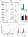
Figure 1
Figure 1. Impact of constitutive NF-κB activation and BLIMP1 deletion on the GC reaction
(A) Left panel, analysis by flow-cytometry of the GC B-cell population (CD19posB220PosCD38LowFasHigh) in spleen, 10 days after primary immunization with SRBC of at least 3 mice per genotype. Right panel, reporter expression in GC B-cells. (B) Analysis of GC B-cells as in A, 21 days after primary immunization with SRBC of at least 3 mice per genotype. (C) Numbers of reporter positive GC B-cells in spleen at day 10 (1° day10) and 21 (1° day21) after primary immunization and of mice re-immunized 30 days after primary immunization and analyzed 10 days thereafter (2° day10). Red bar represents mean of at least 3 mice per genotype. (D) Relative BLIMP1 transcript levels in reporter positive GC B-cells at day 10 after primary immunization. Values represent normalized levels to HPRT, relative to control (Cγ1-cre;eYFPstopFL). Data are represented as mean ±SEM, of 3 mice per genotype. Numbers in graph represent mean value. (E) IgH somatic mutation in reporter positive GC B-cells at day 10 (≥8 sequences per mouse from at least 2 mice per genotype) and day 21 (≥10 sequences per mouse from at least 3 mice per genotype) after primary immunization with SRBC. Red bar represents mean. Average mutation frequency at day 21 is given in red. (F) Relative AICDA transcript levels in reporter positive GC B-cells at day 10 after primary immunization. Values represent normalized levels to HPRT, relative to control (Cγ1-cre;eYFPstopFL). Data are represented as mean ±SEM, of 3 mice per genotype. Numbers in graph represent mean value.
Figure 2
Figure 2. B-cell proliferation and survival upon BLIMP1 deletion and constitutive NF-κB activation
(A) DNA content of reporter positive GC B-cells at day 10 after primary immunization was determined using the Draq5 DNA binding reagent and flow-cytometry. Data are represented as mean ±SEM of 4 mice per genotype. (B) Cre-mediated recombination efficiency, measured by expression of reporter genes, in purified B-cells from compound mutant mice and control, cultured in-vitro in the presence of αCD40 plus IL4. Data are representative of 3 independent experiments. (C) CFSE dilution profile of in-vitro cultured splenic B-cells 5 days after stimulation with αCD40 plus IL4, from mice of the indicated genotypes. Data are representative of 3 independent experiments. (D) Relative BLIMP1 transcript levels in in-vitro cultured splenic B-cells 4 days after stimulation with αCD40 plus IL4, from mice of the indicated genotypes. Values represent normalized levels to HPRT, relative to control (Cγ1-cre;eYFPstopFL). Data are represented as mean ±SEM, of 3 independent experiments. Numbers in graph represent mean values. (E) Frequency of apoptotic cells within in-vitro cultured splenic B-cells 5 days after stimulation with αCD40 plus IL4, from mice of the indicated genotypes. Data are represented as mean ±SEM of 3 independent experiments. (F) % live cells relative to input in in-vitro cultured splenic B-cells at days 3, 4 and 5 after stimulation with αCD40 plus IL4, from mice of the indicated genotypes. % live cells relative to input is calculated based on the cell number of live cells at day 3, 4 and 5 relative to the cell number of live cells at day 0. Data are represented as mean ±SEM of 3 independent experiments.
Figure 3
Figure 3. BLIMP1 inactivation dominantly abrogates plasma cell formation
(A) Left panel, flow-cytometric analysis of splenic plasma cells (B220LowCD138Pos) at day 10 after primary immunization, of at least 3 mice per genotype. Right panel, frequency of reporter positive plasma cells. (B) Reporter positive plasma cell numbers in spleen at day 10, after primary immunization and secondary immunization. Red bar represents mean of at least 3 mice per genotype. (C) SRBC specific IgG1 serum antibodies at day 10 after primary immunization. Data are represented as mean ±SEM, of 3 mice per genotype. (Low ctrl), unimmunized mice. (High ctrl), serum from mice hyperimmunized with SRBC. See also Figure S1.
Figure 4
Figure 4. Survival of mice carrying the Cγ1-cre transgene together with Blimp1F, IKK2castopFL or both alleles
Kaplan–Meier survival graph of mice of the indicated genotypes. (Med. surv), Median survival. (und), undefined.
Figure 5
Figure 5. Constitutive canonical NF-κB signaling promotes plasma cell hyperplasia
(A) Representative picture of spleens from ≥550 day old mice of the indicated genotypes. 6 mice per genotype were analyzed. (B) Absolute number of reporter positive plasma cells (CD19negCD138pos) in spleen and bone marrow of ≥550 day old mice of the indicated genotypes. Red bar represents mean of at least 6 mice per genotype. (C) Representative immunohistochemical staining of spleens from ≥550 day old mice of the indicated genotypes. Upper panel hematoxilin and eosin, middle panel CD138, and lower panel intracellular Ig. 6 mice analyzed per genotype. Scale bar 1000μm, inset, 100μm. (D) Representative serum protein electrophoresis of sera from ≥550 day old mice of the indicated genotypes. The position of albumin and of other components of the serum is indicated. (*) indicates M-spike/s. At least 10 mice per genotype were analyzed. See also Figure S2.
Figure 6
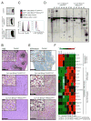
Figure 6. Mice with BLIMP1 deletion alone and in combination with constitutive canonical NF-κB signaling develop lymphoma with an activated B-cell phenotype
(A) Representative pictures of spleens of compound mutant and control mice. (B) Representative hematoxilin and eosin staining of spleens of compound mutant (with 6 Cγ1-cre;Blimp1FFeYFPstopFL, and 5 Cγ1-cre;Blimp1FFIKK2castopFL mice analyzed) and control mice. Scale bar 1000μm, inset, 100μm. (C) Representative analysis of cell size and CD19 expression of reporter positive tumor cells from 6 Cγ1-cre;Blimp1FFeYFPstopFL, and 5 Cγ1-cre;Blimp1FFIKK2castopFL mice and control mice. (D) Southern blot analysis for tumor clonality, using a JH4 probe. Clonal tumors usually exhibit two bands, corresponding to VDJ and DJ rearrangements. (E) Representative immunohistochemical staining for MUM1/IRF4 of spleens of compound mutant (with 5 Cγ1-cre;Blimp1FFeYFPstopFL, and 5 Cγ1-cre;Blimp1FFIKK2castopFL mice analyzed) and control mice. Scale bar 1000μm, inset, 100μm. (F) Heat-map showing the relative transcript levels of candidate genes in lymphoma samples compared to GC B-cells. For heat-map generation, the HPRT-normalized value of each candidate gene was normalized to have mean zero, variance one. See also Figure S3 and Table S1.

Comment in

References

    1. Alizadeh AA, Eisen MB, Davis RE, Ma C, Lossos IS, Rosenwald A, Boldrick JC, Sabet H, Tran T, Yu X, et al. Distinct types of diffuse large B-cell lymphoma identified by gene expression profiling. Nature. 2000;403:503–511. - PubMed
    1. Angelin-Duclos C, Cattoretti G, Lin KI, Calame K. Commitment of B lymphocytes to a plasma cell fate is associated with Blimp-1 expression in vivo. J Immunol. 2000;165:5462–5471. - PubMed
    1. Annunziata CM, Davis RE, Demchenko Y, Bellamy W, Gabrea A, Zhan F, Lenz G, Hanamura I, Wright G, Xiao W, et al. Frequent engagement of the classical and alternative NF-kappaB pathways by diverse genetic abnormalities in multiple myeloma. Cancer Cell. 2007;12:115–130. - PMC - PubMed
    1. Banham AH, Connors JM, Brown PJ, Cordell JL, Ott G, Sreenivasan G, Farinha P, Horsman DE, Gascoyne RD. Expression of the FOXP1 transcription factor is strongly associated with inferior survival in patients with diffuse large B-cell lymphoma. Clin Cancer Res. 2005;11:1065–1072. - PubMed
    1. Bolduc A, Long E, Stapler D, Cascalho M, Tsubata T, Koni PA, Shimoda M. Constitutive CD40L expression on B cells prematurely terminates germinal center response and leads to augmented plasma cell production in T cell areas. J Immunol. 2010;185:220–230. - PMC - PubMed

Publication types